. 2003 Feb;23(4):1418-27.
doi: 10.1128/MCB.23.4.1418-1427.2003.

Regulation of c-Rel nuclear localization by binding of Ca2+/calmodulin

Affiliations

Regulation of c-Rel nuclear localization by binding of Ca2+/calmodulin

Asa Antonsson et al. Mol Cell Biol. 2003 Feb.

Abstract

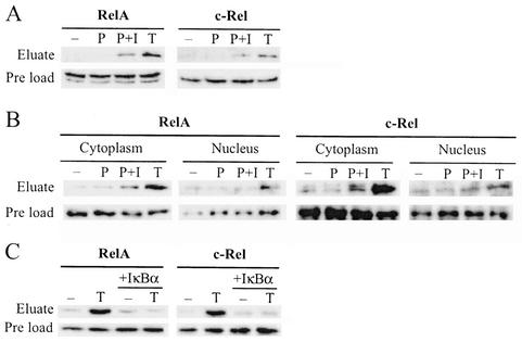
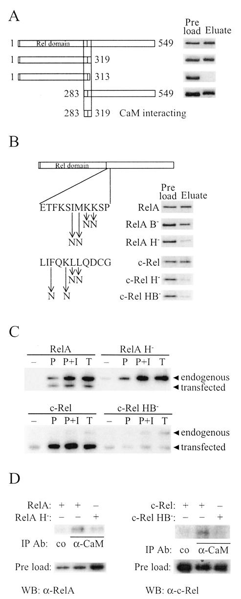
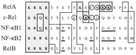
The NF-kappa B/Rel family of transcription factors participates in the control of a wide array of genes, including genes involved in embryonic development and regulation of immune, inflammation, and stress responses. In most cells, inhibitory I kappa B proteins sequester NF-kappa B/Rel in the cytoplasm. Cellular stimulation results in the degradation of I kappa B and modification of NF-kappa B/Rel proteins, allowing NF-kappa B/Rel to translocate to the nucleus and act on its target genes. Calmodulin (CaM) is a highly conserved, ubiquitously expressed Ca(2+) binding protein that serves as a key mediator of intracellular Ca(2+) signals. Here we report that two members of the NF-kappa B/Rel family, c-Rel and RelA, interact directly with Ca(2+)-loaded CaM. The interaction with CaM is greatly enhanced by cell stimulation, and this enhancement is blocked by addition of I kappa B. c-Rel and RelA interact with CaM through a similar sequence near the nuclear localization signal. Compared to the wild-type protein, CaM binding-deficient mutants of c-Rel exhibit increases in both nuclear accumulation and transcriptional activity on the interleukin 2 and granulocyte macrophage colony-stimulating factor promoters in the presence of a Ca(2+) signal. Conversely, for RelA neither nuclear accumulation nor transcriptional activity on these promoters is increased by mutation of the sequence interacting with CaM. Our results suggest that CaM binds c-Rel and RelA after their release from I kappa B and can inhibit nuclear import of c-Rel while letting RelA translocate to the nucleus and act on its target genes. CaM can therefore differentially regulate the activation of NF-kappa B/Rel proteins following stimulation.


Figures

FIG. 1.
FIG. 1.
NF-κB/Rel proteins bind CaM. (A) A calf thymus nuclear protein preparation purified by a series of chromatographic steps including CaM-Sepharose was analyzed by EMSA with a κB sequence used as a probe. A 100-fold molar excess of the same DNA sequence (s) or a non-κB sequence (ns) was added as competitor where indicated. In the first lane (co), no protein was added to the binding reaction. (B) RelA, c-Rel, or NF-κB1 (p50) expressed in vitro in a reticulocyte lysate was incubated with CaM-Sepharose in the presence of Ca2+ (+) or EDTA (−), eluted with 1 M NaCl plus 2 mM EDTA, and analyzed by SDS-PAGE and autoradiography. Preloads (lane 1) represent 40% (RelA and NFκB1) and 20% (c-Rel) of the protein amount incubated with CaM-Sepharose or protein A-Sepharose. RelA and c-Rel binding was inhibited by prebinding a fourfold molar excess of a CaM-binding peptide, amino acids 290 to 309 of CaM kinase II, to the CaM-Sepharose (lane 4), and RelA and c-Rel did not bind to protein A-Sepharose (lanes 5 and 6).
FIG. 2.
FIG. 2.
RelA and c-Rel binding to CaM is increased by NF-κB/Rel-activating stimuli. (A) Jurkat cells were unstimulated (−) or stimulated for 1 h with PMA (P), PMA plus ionomycin (P+I), or TNF-α (T), and whole-cell extracts were prepared. Extracts were incubated with CaM affinity resin, bound proteins were eluted and separated by SDS-PAGE, and RelA and c-Rel were detected by Western blot analysis. Preload lanes contain an equal volume of each extract before incubation with the CaM affinity resin. (B) Cytoplasmic and nuclear extracts of Jurkat cells, stimulated as described for panel A, were analyzed as for panel A. (C) Whole-cell extracts of Jurkat cells, stimulated where indicated for 1 h with TNF-α (T), were incubated with CaM affinity resin in the presence or absence of recombinant IκBα. Eluates were analyzed as for panel A.
FIG. 3.
FIG. 3.
Identification of the CaM binding sequences of RelA and c-Rel. (A) E. coli-produced and purified full-length (amino acids 1 to 549) and the schematically drawn deletion derivatives of RelA were assayed for CaM-Sepharose binding in the presence of Ca2+ as described for Fig. 1B, except that proteins were visualized by silver staining. (B) E. coli-produced RelA or in vitro-translated c-Rel and derivatives with the point mutations in the CaM binding sequences shown to the left were assayed for CaM-Sepharose binding in thepresence of Ca2+ as described for Fig. 1B. RelA was visualized by silver staining and c-Rel by autoradiography. (C) Jurkat cells transiently transfected with vectors encoding RelA, RelA H, c-Rel, or c-Rel HB were unstimulated (−) or stimulated with PMA (P), PMA plus ionomycin (P+I), or TNF-α (T) for 20 min (RelA) or 1 h (c-Rel) prior to harvest. Whole-cell extracts were then incubated with CaM affinity resin, and bound proteins were eluted and separated by SDS-PAGE. Endogenous RelA and c-Rel and transiently expressed proteins from transfected cDNAs were detected by Western blot analysis. (D) DG75 cells were transiently transfected with vectors encoding the indicated NF-κB/Rel proteins, and whole-cell extracts were subjected to immunoprecipitation with a mouse monoclonal α-CaM antibody or a control (co) mouse monoclonal α-Flag M2 antibody. Coimmunoprecipitated RelA or c-Rel was detected by Western blot analysis. Preload lanes contain an equal volume of each extract.
FIG. 4.
FIG. 4.
Mutation of the CaM binding sequences does not affect DNA binding or IκB inhibition properties of RelA or c-Rel. RelA (A) or c-Rel (B) and their respective CaM binding-deficient derivatives were in vitro translated by using a rabbit reticulocyte lysate system and analyzed for DNA binding activity by EMSA with a κB sequence as probe. Where indicated (+), proteins were preincubated with E. coli-produced and purified IκBα before addition of DNA. In the first lane of each gel, no protein was added to the binding reaction. lysate, binding reactions with rabbit reticulocyte lysate without in vitro-translated protein.
FIG. 5.
FIG. 5.
Functional importance of the CaM binding sequences of c-Rel and RelA. c-Rel, c-Rel H, c-Rel HB, RelA, or RelA H were transiently expressed in Jurkat cells together with an IL-2 (A) or GM-CSF (B) reporter plasmid. Twenty-two hours after transfection, cells were stimulated for 2 h with PMA or PMA plus ionomycin. Values shown are the ratios between the reporter gene activity in the presence of ionomycin and the reporter gene activity in the absence of ionomycin and are normalized to empty vector. Bars represent average ratios from at least three independent transfections ± standard errors of the means.
FIG. 6.
FIG. 6.
The nuclear accumulation of c-Rel, but not of RelA, is negatively regulated by its CaM binding site. (A) Jurkat cells were transfected with expression plasmids for c-Rel or the point-mutated derivatives as indicated on the left. Twenty-four hours after transfection, cells were stimulated for 30 min with PMA (P), PMA plus ionomycin (P+I), or TNF-α (Τ). Cytoplasmic (c) and nuclear (n) extracts were prepared and analyzed by Western blotting with the anti-c-Rel C antibody. As a control of the purity of the cytoplasmic and nuclear extracts, the localization of tubulin (cytoplasmic) and that of histone H1 (nuclear) were determined by Western blotting. Shown are tubulin and histone H1 Western blots with extracts from cells transfected with wild-type c-Rel plasmid. (B) Quantitation of the results shown in panel A and two other corresponding Western blot experiments. The ratios of nuclear and cytoplasmic contents of c-Rel ± standard errors of the means (SEM) are shown. (C) DG75 cells were transfected with c-Rel or c-Rel HB expression plasmids and were harvested 24 h later. Analysis of cytoplasmic (c) and nuclear (n) extracts was performed as described for panel A. (D) Jurkat cells were transfected with RelA or RelA H expression plasmids. After 24 h, cells were stimulated as for panel A. Cytoplasmic (c) and nuclear (n) extracts were prepared, and RelA proteins were detected by Western blotting performed with the anti-p65 C-20 antibody. (E) Quantitation of the results shown in panel D and two other corresponding Western blot experiments. The ratios of nuclear and cytoplasmic contents of RelA ± SEM are shown.
FIG. 7.
FIG. 7.
Comparison of the CaM binding sequences of RelA and c-Rel with the corresponding sequences of other mammalian NF-κB/Rel proteins. Sequences surrounding the identified residues important for CaM binding of mouse RelA (amino acids 301 to 319) and c-Rel (amino acids 294 to 312) were aligned with the corresponding sequences of NF-κB1 (amino acids 369 to 387), RelB (amino acids 388 to 406), and the cloned human NF-κB2 (amino acids 338 to 356) by using the ClustalW program (http://www2.ebi.ac.uk/clustalw). Hydrophobic and basic residues are indicated by open and shaded boxes, respectively. Residues of RelA and c-Rel that were mutated to disrupt CaM binding are circled, and potential α-helix-disturbing amino acids, P or rows of G, in the corresponding regions of NF-κB1, NF-κB2, and RelB are underlined.

Similar articles

Cited by

References

    1. Arenzana Seisdedos, F., P. Turpin, M. Rodriguez, D. Thomas, R. T. Hay, J. L. Virelizier, and C. Dargemont. 1997. Nuclear localization of I kappa B alpha promotes active transport of NF-kappa B from the nucleus to the cytoplasm. J. Cell Sci. 110:369-378. - PubMed
    1. Barkett, M., and T. D. Gilmore. 1999. Control of apoptosis by Rel/NF-kappaB transcription factors. Oncogene 18:6910-6924. - PubMed
    1. Beg, A. A., T. S. Finco, P. V. Nantermet, and A. S. Baldwin, Jr. 1993. Tumor necrosis factor and interleukin-1 lead to phosphorylation and loss of I kappa B alpha: a mechanism for NF-kappa B activation. Mol. Cell. Biol. 13:3301-3310. - PMC - PubMed
    1. Bird, T. A., K. Schooley, S. K. Dower, H. Hagen, and G. D. Virca. 1997. Activation of nuclear transcription factor NF-kappaB by interleukin-1 is accompanied by casein kinase II-mediated phosphorylation of the p65 subunit. J. Biol. Chem. 272:32606-32612. - PubMed
    1. Capobianco, A. J., D. L. Simmons, and T. D. Gilmore. 1990. Cloning and expression of a chicken c-rel cDNA: unlike p59v-rel, p68c-rel is a cytoplasmic protein in chicken embryo fibroblasts. Oncogene 5:257-265. - PubMed

Publication types

MeSH terms

LinkOut - more resources