. 2003 Feb;23(4):1441-52.
doi: 10.1128/MCB.23.4.1441-1452.2003.

Direct kinase-to-kinase signaling mediated by the FHA phosphoprotein recognition domain of the Dun1 DNA damage checkpoint kinase

Affiliations

Direct kinase-to-kinase signaling mediated by the FHA phosphoprotein recognition domain of the Dun1 DNA damage checkpoint kinase

Vladimir I Bashkirov et al. Mol Cell Biol. 2003 Feb.

Abstract

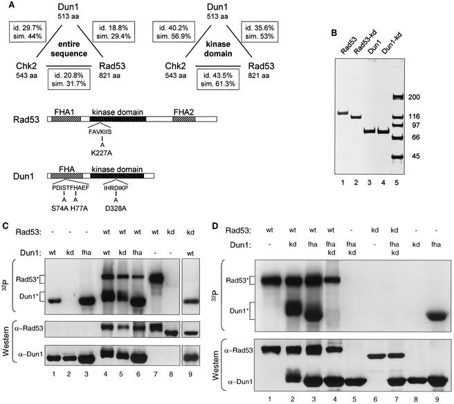
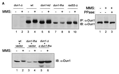
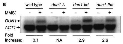
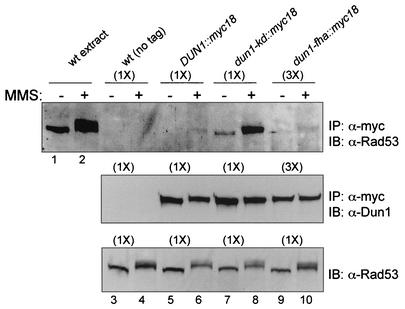
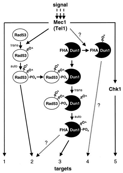
The serine-threonine kinase Dun1 contains a forkhead-associated (FHA) domain and functions in the DNA damage checkpoint pathway of Saccharomyces cerevisiae. It belongs to the Chk2 family of checkpoint kinases, which includes S. cerevisiae Rad53 and Mek1, Schizosaccharomyces pombe Cds1, and human Chk2. Dun1 is required for DNA damage-induced transcription of certain target genes, transient G(2)/M arrest after DNA damage, and DNA damage-induced phosphorylation of the DNA repair protein Rad55. Here we report that the FHA phosphoprotein recognition domain of Dun1 is required for direct phosphorylation of Dun1 by Rad53 kinase in vitro and in vivo. trans phosphorylation by Rad53 does not require the Dun1 kinase activity and is likely to involve only a transient interaction between the two kinases. The checkpoint functions of Dun1 kinase in DNA damage-induced transcription, G(2)/M cell cycle arrest, and Rad55 phosphorylation are severely compromised in an FHA domain mutant of Dun1. As a consequence, the Dun1 FHA domain mutant displays enhanced sensitivity to genotoxic stress induced by UV, methyl methanesulfonate, and the replication inhibitor hydroxyurea. We show that the Dun1 FHA domain is critical for direct kinase-to-kinase signaling from Rad53 to Dun1 in the DNA damage checkpoint pathway.


Figures

FIG. 1.
FIG. 1.
Rad53 kinase directly phosphorylates Dun1 kinase in vitro, dependent on the FHA domain of Dun1. (A) Schematic representation of the relatedness of Rad53, Dun1, and Chk2 kinases and of the Rad53 and Dun1 mutants used in this study. Top panel, Dun1, Rad53, and human Chk2 are related FHA domain kinases. Their entire sequences or their kinase domains were aligned by using the algorithm of Needleman-Wunsch, and the overall sequence identities (id.) and similarities (sim.) are indicated. aa, amino acids. Bottom panel, the rad53-kd allele changes an invariant lysine residue (K227) in subdomain II, which is directly involved in phosphotransfer (22). Rad53-K227A was shown to be nonfunctional and kinase deficient (4, 59). The dun1-kd allele changes an invariant aspartic acid residue (D328) in subdomain VI, which has been implicated in the catalytic mechanism (22). Dun1-D328A was previously shown to be nonfunctional and kinase deficient (25, 61). The Dun1 FHA domain mutant was created by changing serine 74 and histidine 77, two of the four invariable residues in FHA domains (24), to alanine. Previous work with the FHA domain of fission yeast Cds1 showed that such a double mutation abolishes its function (7). The dun1-H77A mutation alone was previously found to have little phenotypic consequence (25). (B) Partial purification of Rad53 and Dun1 kinases. One microgram of purified GST-tagged kinase was analyzed by SDS-4 to 12% PAGE and visualized with Coomassie brilliant blue R250 staining. Wild-type Rad53 (lane 1) and Dun1 (lane 3) proteins were isolated after cell exposure to 0.1% MMS for 2 h, which resulted in an electrophoretic mobility shift caused by phosphorylation (see also panel C and Fig. 2A). The Rad53 (lane 2) and Dun1 (lane 4) kinase-deficient proteins were isolated from cells not exposed to MMS for technical reasons related to protein stability. Control experiments showed no difference between Rad53-kd and Dun1-kd isolated after overexpression from induced or uninduced cells in SDS-PAGE or in in vitro kinase experiments (data not shown). The full-length kinases constituted more than 95% of the preparations as quantified in gel scans. Lane 5, molecular mass standards (in kilodaltons). (C) In vitro phosphorylation of Dun1 kinase by Rad53 kinase. In vitro kinase assays with GST-affinity purified wild-type and mutant proteins (see panel B) were performed. GST-Dun1 fusions were overexpressed in WDHY1413 (dun1-Δ), while all GST-Rad53 fusions were in DES453 (rad53-Δ). Proteins were resolved by SDS-8% PAGE and transferred to nitrocellulose filters. Upper panel, autoradiogram to assess incorporation of 32P into the proteins during the reaction. Rad53* and Dun1* show the positions of the respective phosphorylated protein species. Lower panel, immunoblot analysis of the same filter with anti-Rad53 and anti-Dun1 rat antibodies. wt, wild type; kd, kinase-deficient mutants; fha, FHA domain mutant. Lanes 1 to 8 are from one gel with intervening lanes spliced out; lane 9 is from a different gel. (D) In vitro phosphorylation depends on the Dun1 FHA domain. An in vitro kinase assay was performed with purified GST-fusion proteins overexpressed as described for panel C. Upper panel, 32P incorporation; lower panel, immunoblot analysis with anti-Rad53 and anti-Dun1 rat antibodies. All samples were analyzed on the same gel, but some intervening lanes were spliced out.
FIG. 2.
FIG. 2.
In vivo phosphorylation of Dun1 kinase depends on the Dun1 FHA domain. (A) Dun1 hyperphosphorylation depends on its FHA domain and Rad53 kinase. Upper left panel, immunoprecipitation (IP)-immunoblotting (IB) analysis of Dun1 protein level and phosphorylation status in wild-type (DES460) (lanes 3 and 4), dun1-Δ (MHY26) (lanes 1 and 2), dun1-kd (WDHY1620) (lanes 5 and 6), dun1-fha (WDHY1619) (lanes 7 and 8), and rad53-Δ (DES453) (lanes 9 and 10) strains before and after DNA damage (2 h in 0.1% MMS). Anti-Dun1 rabbit antibodies were used for IP. Immunodetection was performed with rat anti-Dun1 antibodies and the ECL system (Amersham Pharmacia Biotech). Upper right panel, IP mixtures from wild-type cells (DES460) exposed to 0.1% MMS for 2 h (lanes 2 and 3) or left without MMS (lane 1) were either incubated with 1,000 U of λ phosphatase (PPase) (New England Biolabs) at 30°C for 30 min (lane 3) or left untreated (lanes 1 and 2). Dun1 protein was detected as described above. Lower panel, immunoblot of extracts from wild-type (CRY1 with empty vector pYES-TRP1) (lanes 1 and 2), dun1-fha (WDHY1751 with empty vector) (lanes 3 and 4), and dun1-Δ (MHY26 containing plasmid pYES-TRP1-Dun1-fha where Dun1-fha is overexpressed from the GAL1 promoter) (lanes 5 and 6) cells, using rat anti-GST-Dun1 antibodies. Cells were induced for 3 h with 2% galactose and subsequently exposed to 0.1% MMS for 2 h or left without MMS. (B) dun1-fha has no effect on the steady-state and DNA damage-induced DUN1 mRNA levels. Northern blot analysis of DUN1 mRNA levels in wild-type (DES460), dun1-Δ (WDHY1757), dun1-kd (WDHY1620), and dun1-fha (WDHY1619) strains is shown. DUN1 transcript levels in wild-type and mutant strains were determined before and after DNA damage (0.1% MMS for 1 h) and normalized against the ACT1 transcript level. The DNA damage induction of the DUN1 transcript is expressed as fold increase. NA, not applicable.
FIG. 2.
FIG. 2.
In vivo phosphorylation of Dun1 kinase depends on the Dun1 FHA domain. (A) Dun1 hyperphosphorylation depends on its FHA domain and Rad53 kinase. Upper left panel, immunoprecipitation (IP)-immunoblotting (IB) analysis of Dun1 protein level and phosphorylation status in wild-type (DES460) (lanes 3 and 4), dun1-Δ (MHY26) (lanes 1 and 2), dun1-kd (WDHY1620) (lanes 5 and 6), dun1-fha (WDHY1619) (lanes 7 and 8), and rad53-Δ (DES453) (lanes 9 and 10) strains before and after DNA damage (2 h in 0.1% MMS). Anti-Dun1 rabbit antibodies were used for IP. Immunodetection was performed with rat anti-Dun1 antibodies and the ECL system (Amersham Pharmacia Biotech). Upper right panel, IP mixtures from wild-type cells (DES460) exposed to 0.1% MMS for 2 h (lanes 2 and 3) or left without MMS (lane 1) were either incubated with 1,000 U of λ phosphatase (PPase) (New England Biolabs) at 30°C for 30 min (lane 3) or left untreated (lanes 1 and 2). Dun1 protein was detected as described above. Lower panel, immunoblot of extracts from wild-type (CRY1 with empty vector pYES-TRP1) (lanes 1 and 2), dun1-fha (WDHY1751 with empty vector) (lanes 3 and 4), and dun1-Δ (MHY26 containing plasmid pYES-TRP1-Dun1-fha where Dun1-fha is overexpressed from the GAL1 promoter) (lanes 5 and 6) cells, using rat anti-GST-Dun1 antibodies. Cells were induced for 3 h with 2% galactose and subsequently exposed to 0.1% MMS for 2 h or left without MMS. (B) dun1-fha has no effect on the steady-state and DNA damage-induced DUN1 mRNA levels. Northern blot analysis of DUN1 mRNA levels in wild-type (DES460), dun1-Δ (WDHY1757), dun1-kd (WDHY1620), and dun1-fha (WDHY1619) strains is shown. DUN1 transcript levels in wild-type and mutant strains were determined before and after DNA damage (0.1% MMS for 1 h) and normalized against the ACT1 transcript level. The DNA damage induction of the DUN1 transcript is expressed as fold increase. NA, not applicable.
FIG. 3.
FIG. 3.
Rad53 and Dun1 kinases form a complex in vivo. Immunoprecipitation (IP)-immunoblotting (IB) analysis of Rad53- and Dun1-containing complexes is shown. Upper panel, immunoprecipitation of Dun1-myc18 proteins with 9E10 anti-Myc antibodies from the wild type (wt) with untagged DUN1 (CRY1) (lanes 3 and 4), the wild type with DUN1::myc18 (WDHY1934) (lanes 5 and 6), the dun1-kd::myc18 mutant (WDHY1935) (lanes 7 and 8), and the dun1-fha::myc18 mutant (WDHY1936) (lanes 9 and 10), followed by immunoblotting with goat anti-Rad53 antibodies. In lanes 1 and 2, extract from 0.5 optical density unit of wild-type cells (CRY1) was loaded as size standards for activated and nonactivated Rad53. Cells were exposed to 0.1% MMS for 2 h prior to harvesting or not exposed. Note that 250 optical density units of cells was used in lanes 3 to 8 (1×), whereas 750 optical density units was used in lanes 9 and 10 (3×), because the cellular level of the Dun1-fha protein is about threefold lower (see Fig. 2A). Middle panel, loading control for Dun1 levels. The procedure was as for the upper panel, but immunoblotting was with rat anti-Dun antibodies. Lower panel, extract control for Rad53 level. Extracts from 1.5 optical density units of the same cell cultures used for the immunoprecipitation experiment shown in the upper and middle panels were blotted directly with goat anti-Rad53 antibodies. Note that the same amount was used in all lanes.
FIG. 4.
FIG. 4.
The FHA domain is important for the checkpoint function of Dun1 kinase. (A) Scheme for the G2/M arrest assay. G1 phase synchronized cells were released into the cell cycle at the restrictive temperature for cdc13. After S-phase traversal, DNA damage accumulates, leading to a checkpoint-mediated G2/M arrest with medial nuclear division morphology (see text) (modified from reference 16). (B) dun1-fha is defective for the G2/M cell cycle arrest after DNA damage. The cdc13 DUN1 (WDHY1759), cdc13 DUN1 mec1 (WDHY1887), CDC13 DUN1 (DES460), dun1-Δ cdc13 (WDHY1781), dun1-kd cdc13 (WDHY1782), and dun1-fha cdc13 (WDHY1769) strains were used to determine the kinetics and maintenance of the G2/M arrest. (C) dun1-fha is defective in DNA damage-induced gene expression. Northern blot analysis of the RNR2 transcript levels in wild-type (DES460), dun1-Δ (WDHY1757), dun1-kd (WDHY1620), and dun1-fha (WDHY1619) strains is shown. The transcript level of RNR2 mRNA before and after DNA damage (0.1% MMS for 1 h) was normalized against the ACT1 transcript level and is expressed as fold increase after DNA damage. (D) dun1-fha is defective in DNA damage-induced phosphorylation of Rad55 protein. The Rad55 phosphorylation status in wild-type (wt) (DES460) (lanes 1 and 2), dun1-Δ (WDHY1757) (lanes 3 and 4), dun1-fha (WDHY1619) (lanes 5 and 6), and dun1-kd (WDHY1620) (lanes 7 and 8) strains was analyzed by immunoprecipitation (IP) and immunoblotting (IB). Cells were grown either in the absence or in the presence of 0.1% MMS for 2 h.
FIG. 4.
FIG. 4.
The FHA domain is important for the checkpoint function of Dun1 kinase. (A) Scheme for the G2/M arrest assay. G1 phase synchronized cells were released into the cell cycle at the restrictive temperature for cdc13. After S-phase traversal, DNA damage accumulates, leading to a checkpoint-mediated G2/M arrest with medial nuclear division morphology (see text) (modified from reference 16). (B) dun1-fha is defective for the G2/M cell cycle arrest after DNA damage. The cdc13 DUN1 (WDHY1759), cdc13 DUN1 mec1 (WDHY1887), CDC13 DUN1 (DES460), dun1-Δ cdc13 (WDHY1781), dun1-kd cdc13 (WDHY1782), and dun1-fha cdc13 (WDHY1769) strains were used to determine the kinetics and maintenance of the G2/M arrest. (C) dun1-fha is defective in DNA damage-induced gene expression. Northern blot analysis of the RNR2 transcript levels in wild-type (DES460), dun1-Δ (WDHY1757), dun1-kd (WDHY1620), and dun1-fha (WDHY1619) strains is shown. The transcript level of RNR2 mRNA before and after DNA damage (0.1% MMS for 1 h) was normalized against the ACT1 transcript level and is expressed as fold increase after DNA damage. (D) dun1-fha is defective in DNA damage-induced phosphorylation of Rad55 protein. The Rad55 phosphorylation status in wild-type (wt) (DES460) (lanes 1 and 2), dun1-Δ (WDHY1757) (lanes 3 and 4), dun1-fha (WDHY1619) (lanes 5 and 6), and dun1-kd (WDHY1620) (lanes 7 and 8) strains was analyzed by immunoprecipitation (IP) and immunoblotting (IB). Cells were grown either in the absence or in the presence of 0.1% MMS for 2 h.
FIG. 5.
FIG. 5.
The FHA domain is important for the cellular function of Dun1 kinase. Wild-type (DES460), dun1-Δ (WDHY1757), dun1-kd (WDHY1620), and dun1-fha (WDHY1619) strains were used. (A) The dun1-fha strain is sensitive to HU. Serial dilutions of cultures were spotted on YPD plates with or without HU. Plates were photographed after 2 days. (B) The dun1-fha strain is sensitive to MMS. Survival after acute exposure to MMS was determined. The results of one representative experiment are shown. The differences in sensitivity between the strains were highly reproducible. (C) The dun1-fha strain is sensitive to UV. Survival after UVC exposure was determined. The results of one representative experiment are shown. The differences in sensitivity between the strains were highly reproducible.
FIG. 6.
FIG. 6.
Model for checkpoint signaling by S. cerevisiae checkpoint kinases. DNA damage activates Mec1 and Tel1 kinases, which directly and indirectly control a web of secondary kinases (Rad53, Dun1, and Chk1) and effector targets (1 to 5). Kinases involving trans-phosphorylation and autophosphorylation are activated (not shown for all kinases). After Rad53 is activated, one or several phosphorylated residues become a recognition motif for the Dun1 FHA domain. The ensuing transient association between the two kinases leads to trans-phosphorylation of Dun1 by Rad53. Activated Dun1 kinase may have specific effectors (target 3), or target overlap with Rad53 kinase may lead to additional signal amplification (target 2). A postulated Rad53-independent pathway of Dun1 activation is indicated as Mec1/Tel1 controlling Dun1 kinase in a direct or indirect fashion. The mechanism of this pathway is not understood, but it may lead to signal-specific targeting of specific effectors (target 4). For details, see text.

Similar articles

Cited by

References

    1. Aboussekhra, A., J. E. Vialard, D. E. Morrison, M. A. Delatorreruiz, L. Cernakova, F. Fabre, and N. F. Lowndes. 1996. A novel role for the budding yeast RAD9 checkpoint gene in DNA damage-dependent transcription. EMBO J. 15:3912-3922. - PMC - PubMed
    1. Ahn, J.-Y., X. Li, H. L. Davis, and C. E. Canman. 2002. Phosphorylation of threonine 68 promotes oligomerization and autophosphorylation of the Chk2 protein kinase via the forkhead-associated domain. J. Biol. Chem. 277:19389-19395. - PubMed
    1. Alcasabas, A. A., A. J. Osborn, J. Bachant, F. H. Hu, P. J. H. Werler, K. Bousset, K. Furuya, J. F. X. Diffley, A. M. Carr, and S. J. Elledge. 2001. Mrc1 transduces signals of DNA replication stress to activate Rad53. Nat. Cell Biol. 3:958-965. - PubMed
    1. Allen, J. B., Z. Zhou, W. Siede, E. C. Friedberg, and S. J. Elledge. 1994. The SAD1/RAD53 protein kinase controls multiple checkpoints and DNA damage-induced transcription in yeast. Genes Dev. 8:2401-2415. - PubMed
    1. Bashkirov, V. I., J. S. King, E. V. Bashkirova, J. Schmuckli-Maurer, and W. D. Heyer. 2000. DNA repair protein Rad55 is a terminal substrate of the DNA damage checkpoints. Mol. Cell. Biol. 20:4393-4404. - PMC - PubMed

Publication types

MeSH terms