. 2003 Feb;23(4):1460-9.
doi: 10.1128/MCB.23.4.1460-1469.2003.

Maintenance of open chromatin and selective genomic occupancy at the cell cycle-regulated histone H4 promoter during differentiation of HL-60 promyelocytic leukemia cells

Affiliations

Maintenance of open chromatin and selective genomic occupancy at the cell cycle-regulated histone H4 promoter during differentiation of HL-60 promyelocytic leukemia cells

Hayk Hovhannisyan et al. Mol Cell Biol. 2003 Feb.

Abstract

During the shutdown of proliferation and onset of differentiation of HL-60 promyelocytic leukemia cells, expression of the cell cycle-dependent histone genes is downregulated at the level of transcription. To address the mechanism by which this regulation occurs, we examined the chromatin structure of the histone H4/n (FO108, H4FN) gene locus. Micrococcal nuclease, DNase I, and restriction enzymes show similar cleavage sites and levels of sensitivity at the H4/n locus in both proliferating and differentiated HL-60 cells. In contrast, differentiation-related activation of the cyclin-dependent kinase inhibitor p21(cip1/WAF1) gene is accompanied by increased nuclease hypersensitivity. Chromatin immunoprecipitation assays of the H4/n gene reveal that acetylated histones H3 and H4 are maintained at the same levels in proliferating and postproliferative cells. Thus, the chromatin of the H4/n locus remains in an open state even after transcription ceases. Using ligation-mediated PCR to visualize genomic DNase I footprints at single-nucleotide resolution, we find that protein occupancy at the site II cell cycle element is selectively diminished in differentiated cells while the site I element remains occupied. Decreased occupancy of site II is reflected by loss of the site II binding protein HiNF-P. We conclude that H4 gene transcription during differentiation is downregulated by modulating protein interaction at the site II cell cycle element and that retention of an open chromatin conformation may be associated with site I occupancy.


Figures

FIG. 1.
FIG. 1.
Expression of the histone H4/n gene during PMA-induced differentiation of HL-60 cells. (A) Northern blot analysis was performed with total RNA prepared from PMA-treated (differentiated) and untreated (proliferating) HL-60 cells at the indicated times of treatment from two separate experiments. RNAs were hybridized with probes spanning the coding regions of the genes for H4/n and p21. The H4/n probe detects histone H4 mRNAs derived from multiple members of the histone H4 gene family. The ethidium bromide (EthBr)-stained gel is shown as a control for RNA integrity and quantitation. (B) Semiquantitative RT-PCR was performed with the same RNA samples and primers that detect transcripts specific for the H4/n gene. GAPDH was used as an internal control. The right lane (M) shows a 100-bp repeat marker.
FIG. 2.
FIG. 2.
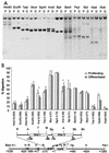
Maintenance of nucleosomal organization at the H4/n gene locus during HL-60 differentiation. (A) Restriction map of the human histone H4/n gene locus and functional organization of the proximal promoter. Arrows show DNA probes used for indirect end labeling to map nuclease accessibility of the H4/n locus. (B to D) Nuclei isolated from control (C) and PMA-treated (PMA) HL-60 cells, as well as naked DNA (N), were incubated with increasing concentrations of MNase for 5 min at room temperature. Purified DNA was digested with HpaI (B), PstI/HindIII (C), and HindIII (D). Southern blots were hybridized to radioactively labeled DNA fragments as follows: Hpa PCR probe (B), XbaI/PstI (C), and HindIII/EcoRI (D). The diagrams at the left of panels B, C, and D indicate the locations of transcription regulatory elements (rectangles), the positions of putative nucleosomes (circles and ovals), and the probes used for hybridization (solid bars). The arrows indicate the transcription start site and length of the protein-coding region. Numbers at the right indicate positions of MNase-sensitive sites.
FIG. 3.
FIG. 3.
Multiple DNase I-hypersensitive sites at the H4/n locus. Nuclei were digested with increasing concentrations of DNase I. DNA was purified and cleaved with PstI/BamHI (A) or HpaI (B). After Southern blotting the filter was hybridized with the XbaI-PstI fragment (A) or the HpaI PCR probe fragment (+336/+500) (B) (see Fig. 2A). The diagram at the left indicates the positions of transcription regulatory elements and the transcription start site. The + sign designates the area of altered nuclease sensitivity. Other symbols and abbreviations are described in the legend to Fig. 2.
FIG. 4.
FIG. 4.
Altered chromatin organization of the p21cip1/WAF1 gene locus during HL-60 cell differentiation. Nuclei isolated from control and PMA-treated (2 days) HL-60 cells were incubated with increasing concentrations of MNase or DNase I for 5 min at room temperature. Purified DNA was digested with StyI, and hypersensitive sites were detected by hybridization with a radiolabeled PCR fragment spanning the p21 promoter region (−1500/−1250). The diagram at the left indicates the position of the transcription start site (arrow) and the probe (bar). Numbers at the right indicate positions of nuclease-sensitive sites relative to the transcription start site of the p21 gene.
FIG. 5.
FIG. 5.
Differences in the restriction enzyme accessibility at the H4/n gene in proliferating and differentiated HL-60 cells. (A) Nuclei were isolated from proliferating (P) and differentiated (D; 2 days of PMA treatment) HL-60 cells and digested with 75 to 500 U of restriction enzyme/ml. Purified DNA was digested to completion with PstI and BamHI. Products were detected by Southern blotting using the XbaI/PstI probe. (B) The relative nuclease sensitivity of restriction enzyme sites was quantitated, and the intensities of radioactive bands were used to calculate the percentage of DNA digested. The relative positions of the restriction enzyme sites are shown in the diagram. The asterisk indicates statistical significance at the P ≤ 0.015 level using the two-tailed paired t test.
FIG. 6.
FIG. 6.
Association of acetylated histones at the histone H4/n locus during HL-60 differentiation. ChIP assays were performed with proliferating HL-60 cells (0) or cells differentiated by treatment with PMA for 1 or 2 days. The antibodies used recognize acetylated histone H4 (α ac. H4) or acetylated histone H3 (α ac. H3). Normal rabbit serum (NRS) was used as a negative control. Precipitated DNA was amplified by PCR using primers detecting specific regions of the H4/n gene, as indicated. A representative set of PCR data from one of two experiments is shown.
FIG. 7.
FIG. 7.
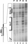
In vivo protein occupancy at the site II cell cycle element of the H4/n gene is selectively downregulated during HL-60 differentiation. Genomic occupancy of the site I and site II regulatory elements of the H4/n gene was analyzed by in vivo DNase I footprinting using LM-PCR. Nuclei from proliferating (P) or differentiated (D) HL-60 cells were digested with DNase I and DNA was purified before LM-PCR analysis. Deproteinized and digested DNA is shown in the right lane (N).
FIG. 8.
FIG. 8.
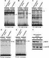
EMSA of protein-DNA interactions at site I and site II of the H4/n promoter. Radiolabeled oligonucleotides detecting the site II binding proteins HiNF-P (A), HiNF-M (B), and HiNF-D (C), the site I binding protein Sp1 (D), and the ubiquitous AP1 factor (E) were each incubated with 4 μg of nuclear protein. Nuclear extracts were prepared from proliferating HL-60 cells (0) or HL-60 cells that were treated with PMA for 15 or 50 h. The same nuclear extracts were used for Western immunoblot analysis (F) to assess HiNF-P protein levels. Lamin B was used as loading control. W, wild-type oligo; M, mutant oligo. Arrows designate specific DNA-protein complexes, and the arrowhead shows free probe.
FIG. 9.
FIG. 9.
Chromatin organization of the H4/n locus. The diagram depicts MNase-sensitive sites (filled triangles), DNase I-hypersensitive sites (open triangles), and restriction enzyme cleavage sites (black vertical bars). Also indicated are the positions of cis regulatory elements (open boxes), the H4/n coding region (hatched box), and the mRNA start site (hooked arrow). The proximal promoter is expanded at the top right to show occupancy of sites I and II (solid box is occupied; dashed box is vacant) in proliferating and differentiated HL-60 cells. Factors known to interact with the H4/n promoter are depicted as ovals. Selective loss of protein-DNA interactions at site II is reflected by downregulation of HiNF-P binding activity and altered sensitivity to cleavage by AvaII (vertical arrow).

Similar articles

Cited by

References

    1. Ausubel, F. M., R. Brent, R. E. Kingston, D. D. Moore, J. G. Seidman, J. A. Smith, and K. Struhl. 1997. Current protocols in molecular biology. John Wiley & Sons, New York, N.Y.
    1. Aziz, F., A. J. van Wijnen, P. S. Vaughan, S. Wu, A. R. Shakoori, J. B. Lian, K. J. Soprano, J. L. Stein, and G. S. Stein. 1998. The integrated activities of IRF-2 (HiNF-M) CDP/cut (HiNF-D) and H4TF-2 (HiNF-P) regulate transcription of a cell cycle controlled human histone H4 gene: mechanistic differences between distinct H4 genes. Mol. Biol. Rep. 25:1-12. - PubMed
    1. Baumbach, L. L., G. S. Stein, and J. L. Stein. 1987. Regulation of human histone gene expression: transcriptional and posttranscriptional control in the coupling of histone messenger RNA stability with DNA replication. Biochemistry 26:6178-6187. - PubMed
    1. Birnbaum, M. J., K. L. Wright, A. J. van Wijnen, A. L. Ramsey-Ewing, M. T. Bourke, T. J. Last, F. Aziz, B. Frenkel, B. R. Rao, N. Aronin, G. S. Stein, and J. L. Stein. 1995. Functional role for Sp1 in the transcriptional amplification of a cell cycle regulated histone H4 gene. Biochemistry 34:7648-7658. - PubMed
    1. Bulger, M., and M. Groudine. 1999. Looping versus linking: toward a model for long-distance gene activation. Genes Dev. 13:2465-2477. - PubMed

Publication types

MeSH terms

LinkOut - more resources