. 2003 Feb;23(4):1470-6.
doi: 10.1128/MCB.23.4.1470-1476.2003.

Disruption of the pelota gene causes early embryonic lethality and defects in cell cycle progression

Affiliations

Disruption of the pelota gene causes early embryonic lethality and defects in cell cycle progression

Ibrahim M Adham et al. Mol Cell Biol. 2003 Feb.

Abstract

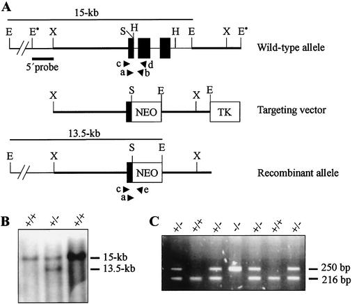
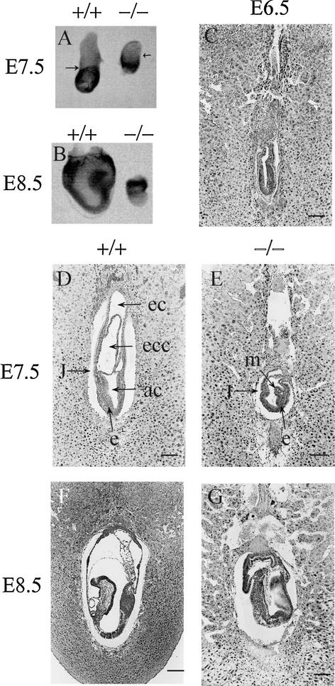
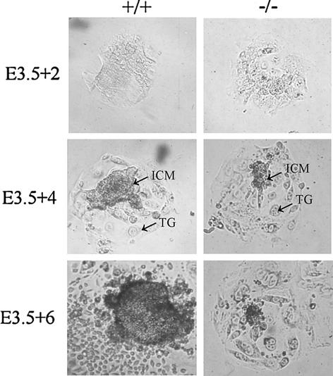
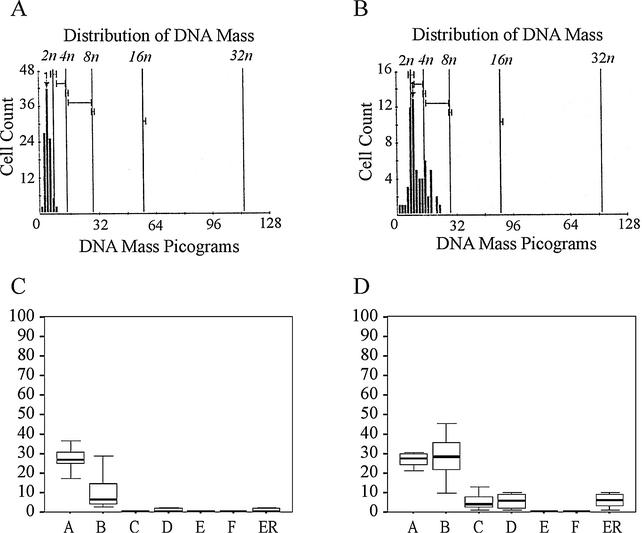
Mutations in either the Drosophila melanogaster pelota or pelo gene or the Saccharomyces cerevisiae homologous gene, DOM34, cause defects of spermatogenesis and oogenesis in Drosophila, and delay of growth and failure of sporulation in yeast. These phenotypes suggest that pelota is required for normal progression of the mitotic and meiotic cell cycle. To determine the role of the pelota in mouse development and progression of cell cycle, we have established a targeted disruption of the mouse PELO: Heterozygous animals are variable and fertile. Genotyping of the progeny of heterozygous intercrosses shows the absence of Pelo(-/-) pups and suggests an embryo-lethal phenotype. Histological analyses reveal that the homozygous Pelo deficient embryos fail to develop past day 7.5 of embryogenesis (E7.5). The failure of mitotic active inner cell mass of the Pelo(-/-) blastocysts to expand in growth after 4 days in culture and the survival of mitotic inactive trophoplast indicate that the lethality of Pelo-null embryos is due to defects in cell proliferation. Analysis of the cellular DNA content reveals the significant increase of aneuploid cells in Pelo(-/-) embryos at E7.5. Therefore, the percent increase of aneuploid cells at E7.5 may be directly responsible for the arrested development and suggests that Pelo is required for the maintenance of genomic stability.


Figures

FIG. 1.
FIG. 1.
Targeted disruption of the Pelo gene. (A) The structures of the wild-type, targeting vector, and recombinant allele are shown together with the relevant restriction sites. The 3.2-kb SpeI-EcoRI fragment containing exons 2 and 3 of the gene was replaced by a pgk-neo selection cassette (NEO). The probe used and the predicted length of EcoRI restriction fragments in Southern blot analysis are shown. Primers F11 (a), R12 (b), F12 (c), R13 (d), and pgk1 (e), used to amplify the wild-type and mutant allele by PCR, are indicated. Abbreviations: TK, Thymidine kinase cassette; E, EcoRI; E*, EcoRI site from polylinker of phage clone; H, HindIII; S, SpeI; X, XbaI. (B) Southern blot analysis of the recombinant ES cell clones. Genomic DNA extracted from ES cell clones was digested with EcoRI and probed with the 5′probe shown in panel A. The wild-type Pelo allele generated a 15-kb EcoRI fragment, whereas the targeted allele yielded a 13.5-kb EcoRI fragment, as indicated in panel A. (C) Second-round PCR products from E3.5 embryos, which were derived from Pelo heterozygous intercrosses and electrophoresed on 2% agarose gel. PCR primers F11 and R12 detect the wild-type allele and generate a 216-bp band; primers F11 and pgk amplify a 250-bp fragment of the targeting allele.
FIG. 2.
FIG. 2.
Morphological and histological analysis of embryos derived from heterozygous intercrosses. (A) Pelo−/− embryos at E7.5 are smaller than wild-type littermates. The arrows point to the junction between the embryonic and extraembryonic regions. (B) E8.5 Pelo−/− embryos have failed to progress and are starting to become resorbed. (C) Histological analysis of E6.5 derived from heterozygous intercrosses does not recognize developmental delay in Pelo−/− embryos. (D) E7.5 wild-type embryos clearly display the distinct germ layers as well as three cavities. (E) E7.5 Pelo−/− embryos are developmentally delayed and have a layer of columnar ectoderm lining the amniotic cavity and a layer of mesodermal cells. The junction between the embryonic and extraembryonic region is clearly seen in the E7.5 mutant embryos. (F) E8.5 wild-type embryo. (G) The E8.5 Pelo−/− embryo is less organized than its wild-type littermates. Abbreviations: ec, ectodermal cavity; ecc, exocoelomic cavity; ac, amniotic cavity; m, mesoderm; e, ectoderm; j, junction between embryonic and extraembryonic regions. (Bar: 120 μm [C to G] or 200 μm [F].)
FIG. 3.
FIG. 3.
Growth of Pelo+/+ and Pelo−/− embryos in culture. Blastocysts (E3.5) derived from heterozygous intercrosses were cultured in vitro in 96-well plates for 7 days and photographed at the 2nd (3.5 + 2), 4th (E3.5 + 4), and 6th day (E3.3 + 6) of culture. TG, trophoplast giant cells.
FIG. 4.
FIG. 4.
Increase of polyploid cells in E7.5 mutant embryos. (A and B) DNA histogram profile of the cells in a wild-type (A) and a developmentally impaired (B) embryo. (C and D) Distribution of the cells with 2N (bar A), 2N to 4N (bar B), 4N (bar C), 4N to 8N (bar D), 8N (bar F), and >5N (bar ER) DNA content in normal (C) and abnormal embryos (D).

References

    1. Auer, G. U., T. O. Caspersson, and A. S. Wallgren. 1980. DNA content and survival in mammary carcinoma. Anal. Quant. Cytol. 2:161-165. - PubMed
    1. Basu, J., B. C. Williams, Z. Li, E. V. Williams, and M. L. Goldberg. 1998. Depletion of a Drosophila homolog of yeast Sup35p disrupts spindle assembly, chromosome segregation, and cytokinesis during male meiosis. Cell Motil. Cytoskeleton 39:286-302. - PubMed
    1. Benachenhou, N., S. Guiral, I. Gorska-Flipot, D. Labuda, and D. Sinnett. 1999. Frequent loss of heterozygosity at the DNA mismatch-repair loci hMLH1 and hMSH3 in sporadic breast cancer. Br. J. Cancer 79:1012-1017. - PMC - PubMed
    1. Brown, E. J., and D. Baltimore. 2000. ATR disruption leads to chromosomal fragmentation and early embryonic lethality. Genes Dev. 14:397-402. - PMC - PubMed
    1. Bunz, F., A. Dutriaux, C. Lengauer, T. Waldman, S. Zhou, J. P. Brown, J. M. Sedivy, K. W. Kinzler, and B. Vogelstein. 1998. Requirement for p53 and p21 to sustain G2 arrest after DNA damage. Science 282:1497-1501. - PubMed

Publication types