

The role of the ubiquitination-proteasome pathway in breast cancer: applying drugs that affect the ubiquitin-proteasome pathway to the therapy of breast cancer
- PMID: 12559038
- PMCID: PMC154126
- DOI: 10.1186/bcr460
The role of the ubiquitination-proteasome pathway in breast cancer: applying drugs that affect the ubiquitin-proteasome pathway to the therapy of breast cancer
Abstract
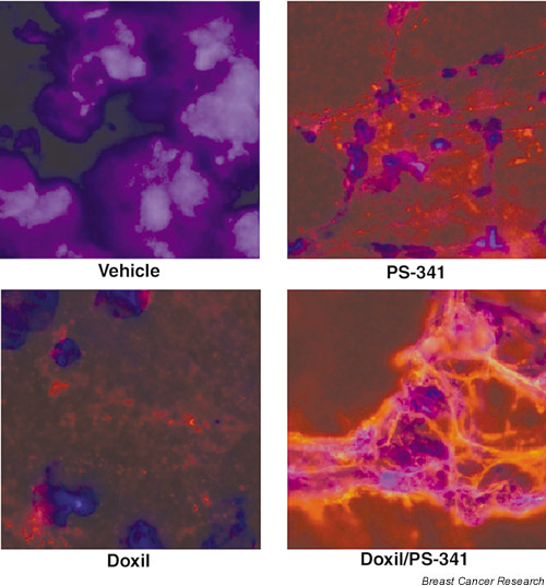
The ubiquitin-proteasome pathway is responsible for most eukaryotic intracellular protein degradation. This pathway has been validated as a target for antineoplastic therapy using both in vitro and preclinical models of human malignancies, and is influenced as part of the mechanism of action of certain chemotherapeutic agents. Drugs whose primary action involves modulation of ubiquitin-proteasome activity, most notably the proteasome inhibitor PS-341, are currently being evaluated in clinical trials, and have already been found to have significant antitumor efficacy. On the basis of the known mechanisms by which these agents work, and the available clinical data, they would seem to be well suited for the treatment of breast neoplasms. Such drugs, alone and especially in combination with current chemotherapeutics, may well represent important advances in the therapy of patients with breast cancer.
Figures

References
-
- Orlowski RZ, Eswara JR, Lafond-Walker A, Grever MR, Orlowski M, Dang CV. Tumor growth inhibition induced in a murine model of human Burkitt's lymphoma by a proteasome inhibitor. Cancer Res. 1998;58:4342–4348. - PubMed
-
- Adams J, Palombella VJ, Sausville EA, Johnson J, Destree A, Lazarus DD, Maas J, Pien CS, Prakash S, Elliott PJ. Proteasome inhibitors: a novel class of potent and effective antitumor agents. Cancer Res. 1999;59:2615–2622. - PubMed
-
- Orlowski RZ, Stinchcombe TE, Mitchell BS, Shea TC, Baldwin AS, Stahl S, Adams J, Esseltine DL, Elliott PJ, Pien CS, Guerciolini R, Anderson JK, Depcik-Smith ND, Bhagat R, Lehman MJ, Novick SC, O'Connor OA, Soignet SL. Phase I trial of the proteasome inhibitor PS-341 in patients with refractory hematologic malignancies. J Clin Oncol. 2002. - PubMed
-
- Kiyomiya K, Matsuo S, Kurebe M. Mechanism of specific nuclear transport of adriamycin: the mode of nuclear translocation of adriamycin-proteasome complex. Cancer Res. 2001;61:2467–2471. - PubMed
-
- Yoshida H, Kitamura K, Tanaka K, Omura S, Miyazaki T, Hachiya T, Ohno R, Naoe T. Accelerated degradation of PML-retinoic acid receptor α (PML-RARA) oncoprotein by all-trans-retinoic acid in acute promyelocytic leukemia: possible role of the proteasome pathway. Cancer Res. 1996;56:2945–2948. - PubMed
Publication types
MeSH terms
Substances
LinkOut - more resources
Full Text Sources
Other Literature Sources
Medical
