

Review
doi: 10.1016/S0165-6147(02)00038-X.
Characterization of the UDP-glucose receptor (re-named here the P2Y14 receptor) adds diversity to the P2Y receptor family
Affiliations
- PMID: 12559763
- PMCID: PMC8653507
- DOI: 10.1016/S0165-6147(02)00038-X
Item in Clipboard
Review
Characterization of the UDP-glucose receptor (re-named here the P2Y14 receptor) adds diversity to the P2Y receptor family
Trends Pharmacol Sci.
2003 Feb.
Abstract
The cloning of a human G-protein-coupled receptor (GPCR) that specifically responds to UDP-glucose and related sugar-nucleotides has been reported recently. This receptor has important structural similarities to known members of the P2Y receptor family but also shows a distinctly different pharmacological response profile. Here, the IUPHAR Subcommittee for P2Y receptor nomenclature and classification review the current knowledge of this receptor and present their reasons for including this receptor in the P2Y receptor family as the P2Y(14) receptor.
Figures
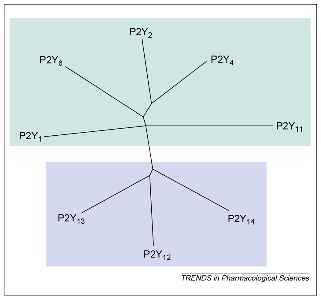
A phylogenetic tree (dendrogram) showing the relationships among the current members of the P2Y receptor family (human P2Y1 P2Y2, P2Y4, P2Y6, P2Y11, P2Y12 and P2Y13 receptors) and the human UDP-glucose receptor (here indicated as the P2Y14 receptor). The P2Y receptors can be divided into two subgroups shown with green and blue backgrounds. Sequences were aligned using clustalx and the tree was built using the treeview software.
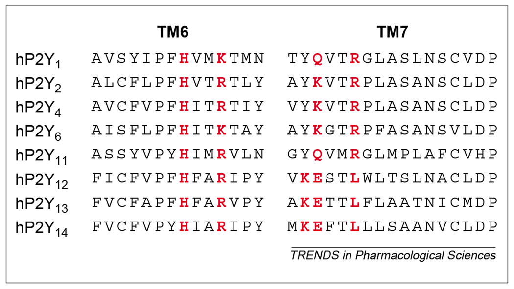
Alignment of putative nucleotide binding motifs in transmembrane domain 6 (TM6) and TM7 of human (h) P2Y receptors. All receptor subtypes share in TM6 the presence of the H and R/K amino acid residues proposed to be crucial for receptor activity. In P2Y1 P2Y2, P2Y4, P2Y6 and P2Y11 receptors, a Y-Q/K-X-X-R motif in TM7 has also been proposed to participate in ligand binding. In P2Y12 and P2Y13 receptors and in the UDP-glucose receptor (here indicated as the P2Y14 receptor subtype), this motif is substituted with K-E-X-X-L. Crucial amino acids for nucleotide binding are highlighted in red. Sequences were aligned using clustalx .
References
-
- Chambers JK et al. (2000) A G protein-coupled receptor for UDP-glucose. J. Biol. Chem 275, 10767–10771 - PubMed
-
- Neary JT et al. (1996) Trophic actions of extracellular nucleotides and nucleosides on glial and neuronal cells. Trends Neurosci. 19, 13–18 - PubMed
-
- Ralevic V and Burnstock G (1998) Receptors for purines and pyrimidines. Pharmacol. Rev 60, 413–492 - PubMed
-
- Burnstock G and Kennedy C (1985) Is there a basis for distinguishing two types of P2-purinoceptor? Gen. Pharmacol 16, 433–440 - PubMed
-
- Abbracchio MP and Burnstock G (1994) Purinoceptors: are there families of P2X and P2Y purinoceptors? Pharmacol. Ther 64, 445–475 - PubMed
Publication types
MeSH terms
Substances
Grants and funding
LinkOut - more resources
Full Text Sources
Other Literature Sources
Molecular Biology Databases
