. 2003 Feb 1;31(3):893-8.
doi: 10.1093/nar/gkg188.

Sir2p suppresses recombination of replication forks stalled at the replication fork barrier of ribosomal DNA in Saccharomyces cerevisiae

Affiliations

Sir2p suppresses recombination of replication forks stalled at the replication fork barrier of ribosomal DNA in Saccharomyces cerevisiae

Alberto Benguría et al. Nucleic Acids Res. .

Abstract

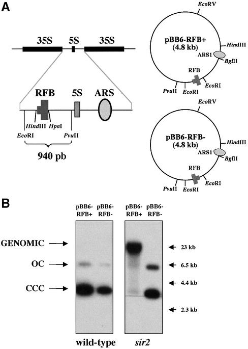
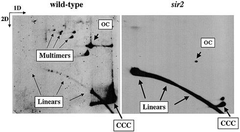
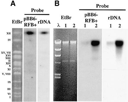
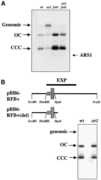
In the ribosomal DNA (rDNA) of Saccharomyces cerevisiae replication forks progressing against transcription stall at a polar replication fork barrier (RFB) located close to and downstream of the 35S transcription unit. Forks blocked at this barrier are potentially recombinogenic. Plasmids bearing the RFB sequence in its active orientation integrated into the chromosomal rDNA in sir2 mutant cells but not in wild-type cells, indicating that the histone deacetylase silencing protein Sir2 (Sir2p), which also modulates the aging process in yeast, suppresses the recombination competence of forks blocked at the rDNA RFB. Orientation of the RFB sequence in its inactive course or its abolition by FOB1 deletion avoided plasmid integration in sir2 mutant cells, indicating that stalling of the forks in the plasmid context was required for recombination to take place. Altogether these results strongly suggest that one of the functions of Sir2p is to modulate access of the recombination machinery to the forks stalled at the rDNA RFB.


Figures

Figure 1
Figure 1
The different behavior of pBB6-RFB+ in sir2 cells. (A) Schematic representation of two rDNA repeats. The 940 bp EcoRI–PvuII fragment, which contains the RFB, was inserted at the EcoRI site of pBB6 in both orientations to yield pBB6-RFB+ and pBB6-RFB–. (B) Stability of pBB6-RFB+ and pBB6-RFB– in wild-type (CT711) and sir2 (sir2Δ::LEU2) strains. Undigested yeast DNA was electrophoresed and subjected to Southern blot hybridization using radiolabeled pBR322 as a probe. Arrows on the right-hand side point to λ-HindIII size markers. A high molecular weight band that co-migrated with genomic DNA was detected only in the case of pBB6-RFB+ isolated from sir2 cells.
Figure 2
Figure 2
pBB6-RFB+ integrated into the genome of sir2 cells. Undigested DNA from wild-type (CT711) and sir2 (sir2Δ::LEU2) strains were subjected to 2D agarose gel electrophoresis and Southern blot hybridization using radiolabeled pBR322 as a probe. Monomeric and multimeric extra-chromosomal forms of pBB6-RFB+ were clearly detected in wild-type cells. In sir2 mutants, however, the only prominent signal corresponded to sheared linear forms.
Figure 3
Figure 3
In sir2 cells pBB6-RFB+ integrated into the rDNA locus at chromosome XII. (A) Undigested DNA from sir2 cells transformed with pBB6-RFB+ was subjected to pulsed field electrophoresis. The gel was stained with ethidium bromide (EtBr lane), to separate the different yeast chromosomes (Roman numerals on the left-hand side) and subjected to Southern blot hybridization using pBR322 as probe (pBB6-RFB+ lane). The stripped blot was re-hybridized with a yeast rDNA probe (rDNA lane). Note that both probes hybridized to a single band that co-migrated with chromosome XII. (B) Yeast DNA obtained from sir2 cells transformed with pBB6-RFB+ was subjected to ultracentrifugation in a CsCl gradient in the presence of Hoescht 33258. Two fractions enriched for bulk DNA (lanes designated 1) or rDNA (lanes designated 2) were isolated from the CsCl gradient, subjected to electrophoresis and hybridized using pBR322 (pBB6-RFB+) or a yeast rDNA fragment (rDNA) as probes. The EtBr panel corresponds to the gel stained with ethidium bromide (λ = lambda/HindIII size marker). In both cases probes hybridized mainly to the rDNA-enriched sample.
Figure 4
Figure 4
(A) Deletion of FOB1 avoided integration of pBB6-RFB+ in sir2 cells. DNA isolated from wild-type (CT711), sir2 (sir2Δ::LEU2), fob1 (fob1Δ::HIS3) and sir2 fob1 double mutants transformed with pBB6-RFB+ were digested with XhoI and XbaI and subjected to electrophoresis and Southern blot hybridization. To avoid cross-hybridization with bacterial sequences, a chromosomal 237 bp HindIII–BglII ARS1 fragment was used as probe. The restriction enzymes used do not cut pBB6-RFB+. A signal co-migrating with genomic DNA was detected only in sir2 cells. The weak band running faster than CCCs corresponded to the endogenous ARS1 fragment (ARS1). (B) Elimination of part of the region downstream of the HpaI site avoided integration in sir2 cells. The maps on top depict the most relevant features of the rDNA RFB and adjacent sequences in pBB6-RFB+ and pBB6-RFB+(del). pBB6-RFB+(del) was obtained as described in Material and Methods and used to transform both wild-type (CT711) and sir2 (sir2Δ::LEU2) strains. Undigested DNA was subjected to electrophoresis and Southern blot hybridization using pBR322 as probe. No hybridization signal was observed co-migrating with genomic DNA.

References

    1. Hernández P., Martín-Parras,L., Martínez-Robles,M.L. and Schvartzman,J.B. (1993) Conserved features in the mode of replication of eukaryotic ribosomal RNA genes. EMBO J., 12, 1475–1485. - PMC - PubMed
    1. Bastia D. and Mohanty,B.K. (1996) Mechanisms for completing DNA replication. In DePamphilis,M.L. (ed.), DNA Replication in Eukaryotic Cells. Cold Spring Harbor Laboratory Press, New York, NY, pp. 177–215.
    1. Hill T.M., Pelletier,A.J., Tecklenburg,M.L. and Kuempel,P.L. (1988) Identification of the DNA sequence from E. coli terminus region that halts replication forks. Cell, 55, 459–466. - PubMed
    1. Brewer B.J. (1988) When polymerases collide: replication and the transcriptional organization of the E. coli chromosome. Cell, 53, 679–686. - PubMed
    1. Olavarrieta L., Hernández,P., Krimer,D.B. and Schvartzman,J.B. (2002) DNA knotting caused by head-on collision of transcription and replication. J. Mol. Biol., 322, 1–6. - PubMed

Publication types

MeSH terms