

Functional and Vbeta repertoire characterization of human CD8+ T-cell subsets with natural killer cell markers, CD56+ CD57- T cells, CD56+ CD57+ T cells and CD56- CD57+ T cells
- PMID: 12562330
- PMCID: PMC1782888
- DOI: 10.1046/j.1365-2567.2003.01575.x
Functional and Vbeta repertoire characterization of human CD8+ T-cell subsets with natural killer cell markers, CD56+ CD57- T cells, CD56+ CD57+ T cells and CD56- CD57+ T cells
Abstract
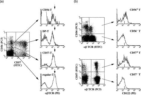
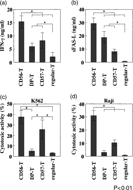
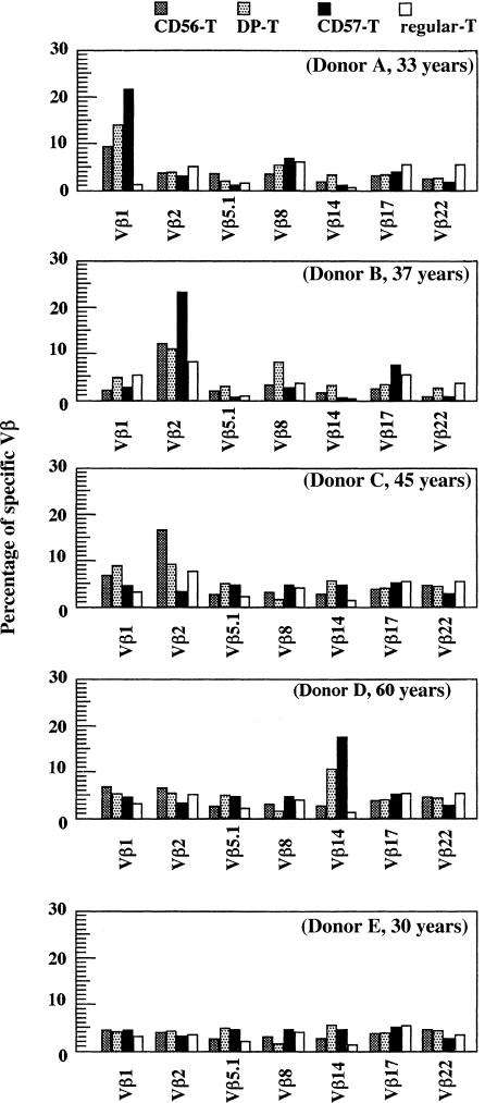
We investigated the individual CD8+ populations with natural killer (NK) cell markers (NK-type T cell); CD56 single positive (CD56)-T cells, CD56/CD57 double positive (DP)-T cells and CD57 single positive (CD57)-T cells in the peripheral blood. All NK-type T-cell populations expressed CD122 and intermediate levels of T-cell receptor (TCR; regular CD8+ T cells are CD122- and express high levels of TCR). The number of both DP-T cells and CD57-T cells, but not CD56-T cells, gradually increased with age. All NK-type T-cell populations produced larger amounts of interferon-gamma than did regular CD8+ T cells after stimulation with interleukin (IL)-2, IL-12 and IL-15. However, CD56-T cells and CD57-T cells but not DP-T cells showed a potent antitumour cytotoxity to NK-sensitive K562 cells, whereas only CD56-T cells showed a potent cytotoxity to NK-resistant Raji cells. Furthermore, although NK-type T cells produced large amounts of soluble Fas-ligands, their cytotoxic activities appeared to be mediated by the perforin/granzyme pathway. The oligoclonal or pauciclonal expansions of certain VbetaT cells were found in each NK-type T-cell population. The non-variant CDR3 region(s) for the TCRbeta chain(s) showed CD57-T cells and CD56-T cells to be derived from distinct origins, while the DP-T cell population consisted of a mixture of the clones seen in both CD56-T cells and CD57-T cells. Our results suggest that CD57-T cells and CD56-T cells are functionally and ontogenically different populations while DP-T cells appear to originate from both CD56-T cells and CD57-T cells.
Figures
Similar articles
-
Characterization of bronchoalveolar lavage T cell subsets in sarcoidosis on the basis of CD57, CD4 and CD8.Clin Exp Immunol. 2003 Sep;133(3):438-47. doi: 10.1046/j.1365-2249.2003.02228.x. Clin Exp Immunol. 2003. PMID: 12930372 Free PMC article.
-
Systematic characterization of human CD8+ T cells with natural killer cell markers in comparison with natural killer cells and normal CD8+ T cells.Immunology. 2001 Jul;103(3):281-90. doi: 10.1046/j.1365-2567.2001.01248.x. Immunology. 2001. PMID: 11454057 Free PMC article.
-
Activation of human T cells with NK cell markers by staphylococcal enterotoxin A via IL-12 but not via IL-18.Clin Exp Immunol. 2002 Jun;128(3):453-9. doi: 10.1046/j.1365-2249.2002.01854.x. Clin Exp Immunol. 2002. PMID: 12109440 Free PMC article.
-
Proportion of Cytotoxic Peripheral Blood Natural Killer Cells and T-Cell Large Granular Lymphocytes in Recurrent Miscarriage and Repeated Implantation Failure: Case-Control Study and Meta-analysis.Arch Immunol Ther Exp (Warsz). 2019 Aug;67(4):225-236. doi: 10.1007/s00005-019-00546-5. Epub 2019 May 30. Arch Immunol Ther Exp (Warsz). 2019. PMID: 31147723 Review.
-
The dual-functional capability of cytokine-induced killer cells and application in tumor immunology.Hum Immunol. 2015 May;76(5):385-91. doi: 10.1016/j.humimm.2014.09.021. Epub 2014 Oct 8. Hum Immunol. 2015. PMID: 25305457 Review.
Cited by
-
Activation of CD11b+ Kupffer cells/macrophages as a common cause for exacerbation of TNF/Fas-ligand-dependent hepatitis in hypercholesterolemic mice.PLoS One. 2013;8(1):e49339. doi: 10.1371/journal.pone.0049339. Epub 2013 Jan 23. PLoS One. 2013. PMID: 23372642 Free PMC article.
-
Roles of Natural Killer T Cells and Natural Killer Cells in Kidney Injury.Int J Mol Sci. 2019 May 20;20(10):2487. doi: 10.3390/ijms20102487. Int J Mol Sci. 2019. PMID: 31137499 Free PMC article. Review.
-
Analysis of the susceptibility of CD57 T cells to CD3-mediated apoptosis.Clin Exp Immunol. 2005 Feb;139(2):268-78. doi: 10.1111/j.1365-2249.2004.02687.x. Clin Exp Immunol. 2005. PMID: 15654825 Free PMC article.
-
Characterization of bronchoalveolar lavage T cell subsets in sarcoidosis on the basis of CD57, CD4 and CD8.Clin Exp Immunol. 2003 Sep;133(3):438-47. doi: 10.1046/j.1365-2249.2003.02228.x. Clin Exp Immunol. 2003. PMID: 12930372 Free PMC article.
-
New insights on the role of CD8(+)CD57(+) T-cells in cancer.Oncoimmunology. 2012 Sep 1;1(6):954-956. doi: 10.4161/onci.20307. Oncoimmunology. 2012. PMID: 23162769 Free PMC article.
References
-
- Abo T, Balch CM. A differentiation antigen of human NK and K cells identified by a monoclonal antibody (HNK-1) J Immunol. 1981;127:1024. - PubMed
-
- Lanier LL, Le AM, Civin CI, Loken MR, Phillips JH. The relationship of CD16 (Leu-11) and Leu-19 (NKH-1) antigen expression on human peripheral blood NK cells and cytotoxic T lymphocytes. J Immunol. 1986;136:4480. - PubMed
-
- Abo T, Watanabe H, Iiai T, Kimura M, Ohtsuka K, Sato K, et al. Extrathymic pathways of T-cell differentiation in the liver and other organs. Int Rev Immunol. 1994;11:61. - PubMed
-
- Lanier LL, Le AM, Phillips JH, Warner NL, Babcock GF. Subpopulations of human natural killer cells defined by expression of the Leu-7 (HNK-1) and Leu-11 (NK-15) antigens. J Immunol. 1983;131:1789. - PubMed
MeSH terms
Substances
LinkOut - more resources
Full Text Sources
Research Materials
Miscellaneous
