. 2003 Feb;131(2):324-34.
doi: 10.1046/j.1365-2249.2003.02053.x.

Monocyte-derived cytokines in multiple sclerosis

Affiliations

Monocyte-derived cytokines in multiple sclerosis

L G Filion et al. Clin Exp Immunol. 2003 Feb.

Abstract

MS is an inflammatory, presumably autoimmune, disease mediated by the activation of T cells, B cells and monocytes (MO). Inflammation is thought to occur early during the relapsing-remitting phase of MS (RRMS), whereas in the later phases of MS such as secondary progressive MS (SPMS), inflammation tends to diminish. Our objective was to compare the types and amounts of proinflammatory and regulatory cytokines produced by MO from relapsing-remitting patients with or without treatment with IFN-beta (RRMS+ therapy, RRMS- therapy), respectively, from secondary progressive patients (SPMS) and from healthy controls (HC). MO were isolated by a density-gradient technique and three different techniques (RNase protection assay, ELISA and intracellular cytokine staining) were used to assess cytokine levels. An increase in IL6, IL12 and TNF-alpha was observed by all three methods for RRMS- therapy and for SPMS patients compared to HC and RRMS+ therapy patients. We conclude that proinflammatory and regulatory monokines can be derived from MO of MS patients and that these levels are modulated by IFN-beta therapy. Although it is believed that inflammation tends to diminish in SPMS patients, our data show that inflammatory cytokines continue to be released at high levels, suggesting that IFN-beta or IL10 treatment may be beneficial for this group.


Figures
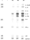
Fig. 1
Fig. 1
Cytokine mRNA levels of MO from MS patients. MO were isolated from peripheral blood and the basal cytokine mRNA levels after 24 h of culture were measured using an RNA protection assay. The figure is representative of the data that were obtained for five experiments. The first and second columns are the positive and negative controls, respectively, provided by the kit. The image was obtained using a phosphorimager.
Fig. 2
Fig. 2
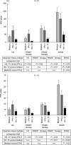
Cytokine mRNA levels of MO from MS patients with or without culture treatment with IFN-β or rhIL-10. MO were isolated from peripheral blood from HC or MS patients and were treated with medium, IFN-β (100 IU/ml) or rhIL-10 (1 ng/ml) for 24 h. The cytokine mRNA levels of the cytokines were measured using an RNA protection assay. Data was obtained from HC (n = 9), RRMS therapy (n = 10), RRMS+ therapy (n = 6) and SPMS (n = 10). The mean and standard deviation of the data are shown.
Fig. 2
Fig. 2
Cytokine mRNA levels of MO from MS patients with or without culture treatment with IFN-β or rhIL-10. MO were isolated from peripheral blood from HC or MS patients and were treated with medium, IFN-β (100 IU/ml) or rhIL-10 (1 ng/ml) for 24 h. The cytokine mRNA levels of the cytokines were measured using an RNA protection assay. Data was obtained from HC (n = 9), RRMS therapy (n = 10), RRMS+ therapy (n = 6) and SPMS (n = 10). The mean and standard deviation of the data are shown.
Fig. 3
Fig. 3
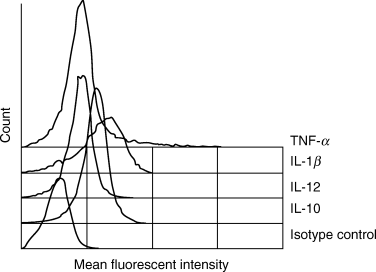
Intracellular cytokine assay. MO from HC or patients were cultured for 24 h in the presence or absence of IFN-β (100 IU/ml) or rhIL-10 (1 ng/ml). Representative data from an SPMS MO cultured in medium are shown. Data were obtained from HC (n = 9), RRMS-therapy (n = 10), RRMS+ therapy (n = 6) and SPMS (n = 10). The isotype control used was the anti-IL2 antibody from Pharmingen.
Fig. 4
Fig. 4
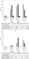
Secreted levels of cytokines of MO from MS patients with or without treatment with IFN-β or rhIL-10. MO were isolated from peripheral blood from HC or MS patients and were treated with medium, IFN-β (100 IU/ml) or rhIL-10 (1 ng/ml) for 24 h. The secreted levels of the cytokines were measured by ELISA. Data were obtained from HC (n = 9), RRMS therapy (n = 10), RRMS+ therapy (n = 6) and SPMS (n = 10) patients. The mean and standard deviation of the data are shown.
Fig. 4
Fig. 4
Secreted levels of cytokines of MO from MS patients with or without treatment with IFN-β or rhIL-10. MO were isolated from peripheral blood from HC or MS patients and were treated with medium, IFN-β (100 IU/ml) or rhIL-10 (1 ng/ml) for 24 h. The secreted levels of the cytokines were measured by ELISA. Data were obtained from HC (n = 9), RRMS therapy (n = 10), RRMS+ therapy (n = 6) and SPMS (n = 10) patients. The mean and standard deviation of the data are shown.

References

    1. Lassmann H, Suchanek G, Ozawa K. Histopathology and the blood–cerebrospinal fluid barrier in multiple sclerosis. Ann Neurol. 1994;36(Suppl.):S42–S46. - PubMed
    1. Ozawa K, Suchanek G, Breitschopf H, et al. Patterns of oligodendroglia pathology in multiple sclerosis. Brain. 1994;117:1311–22. - PubMed
    1. Kouwenhoven M, Teleshova N, Ozenci V, Press R, Link H. Monocytes in multiple sclerosis: phenotype and cytokine profile. J Neuroimmunol. 2001;112:197–205. - PubMed
    1. Calabresi PA, Fields NS, Farnon EC, et al. ELI-spot of Th-1 cytokine secreting PBMC's in multiple sclerosis: correlation with MRI lesions. J Neuroimmunol. 1998;85:212–9. - PubMed
    1. Calabresi PA, Tranquill LR, McFarland HF, Cowan EP. Cytokine gene expression in cells derived from CSF of multiple sclerosis patients. J Neuroimmunol. 1998;89:198–205. - PubMed

Publication types

MeSH terms