. 2003 Feb;84(4):655-68.
doi: 10.1046/j.1471-4159.2003.01571.x.

Palmitic and stearic fatty acids induce caspase-dependent and -independent cell death in nerve growth factor differentiated PC12 cells

Affiliations

Palmitic and stearic fatty acids induce caspase-dependent and -independent cell death in nerve growth factor differentiated PC12 cells

Joel E Ulloth et al. J Neurochem. 2003 Feb.

Abstract

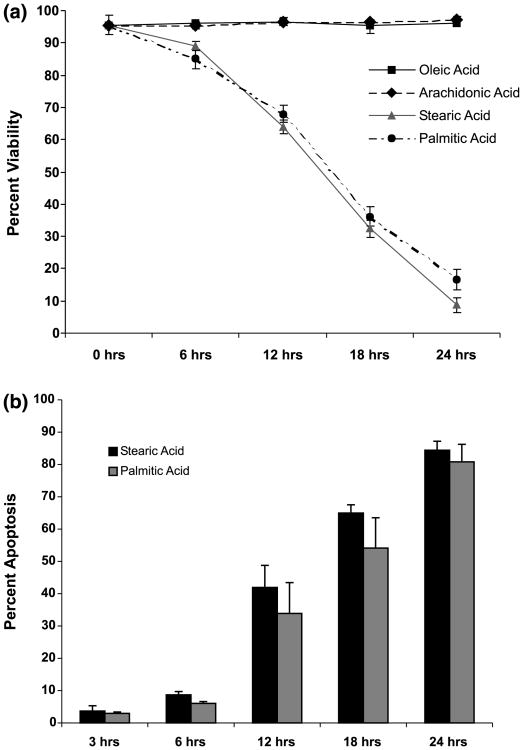
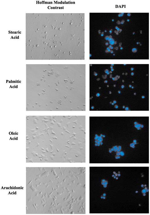
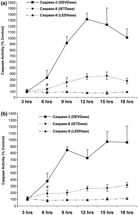
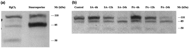
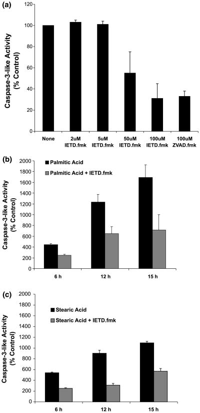
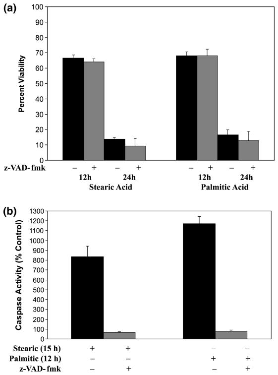
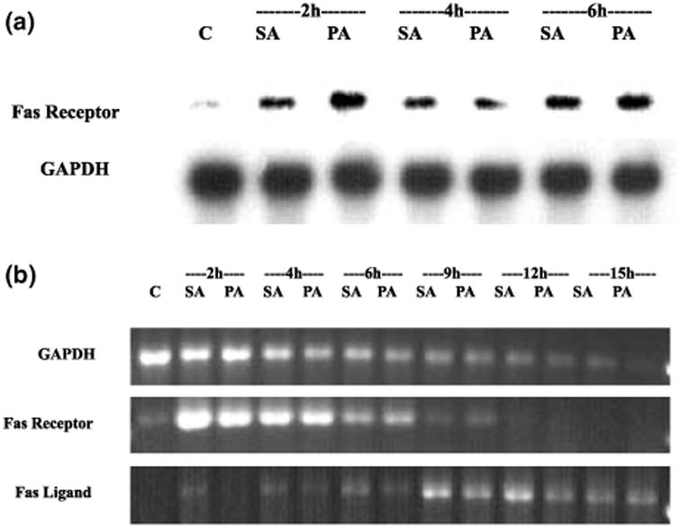
Apoptotic cell death has been proposed to play a role in the neuronal loss observed following traumatic injury in the CNS and PNS. The present study uses an in vitro tissue culture model to investigate whether free fatty acids (FFAs), at concentrations comparable to those found following traumatic brain injury, trigger cell death. Nerve growth factor (NGF)-differentiated PC12 cells exposed to oleic and arachidonic acids (2 : 1 ratio FFA/BSA) showed normal cell survival. However, when cells were exposed to stearic and palmitic acids, there was a dramatic loss of cell viability after 24 h of treatment. The cell death induced by stearic acid and palmitic acid was apoptotic as assessed by morphological analysis, and activation of caspase-8 and caspase-3-like activities. Western blotting showed that differentiated PC12 cells exposed to stearic and palmitic acids exhibited the signature apoptotic cleavage fragment of poly (ADP-ribose) polymerase (PARP). Interestingly, blockade of caspase activities with the pan-caspase inhibitor z-VAD-fmk failed to prevent the cell death observed induced by palmitic or stearic acid. RT-PCR and RNA blot experiments showed an up-regulation of the Fas receptor and ligand mRNA. These findings are consistent with our hypothesis that FFAs may play a role in the cell death associated with trauma in the CNS and PNS.


Figures

Fig. 1
Fig. 1
Stearic and palmitic acid treatments result in loss of viability and increased apoptosis. (a) Stearic and palmitic acid treatments (300 μm) result in loss of viability of NGF-differentiated PC12 cells. Differentiated PC12 cells were treated for up to 24 h with either stearic, palmitic, oleic or arachidonic acid complexed with BSA (2 : 1 ratio). Viability was assessed by trypan blue exclusion during the course of fatty acid treatment. (b) Stearic and palmitic acid treatments increase the percentage of nuclei exhibiting apoptotic morphology. Cells were fixed and stained with 4′,6′-diamidino-2-phenylindole (DAPI) during the course of stearic and palmitic acid treatments. Nuclei were visualized under fluorescent microscopy and nuclei exhibiting condensed and/or fragmented morphology were scored as apoptotic. A minimum of 1000 cells were counted per sample for viability and apoptosis assays. Data represent the mean ± SD of three independent experiments performed in triplicate.
Fig. 2
Fig. 2
Hoffmann modulation contrast and fluorescent micrographs of NGF-differentiated PC12 cells exposed to selected fatty acids complexed with BSA (2 : 1). Images show NGF-differentiated PC12 cells exposed to the indicated fatty acid for 24 h. Fluorescent nuclear staining with DAPI was performed as described in Materials and methods. Panels on the left show Hoffman modulation contrast images of cells before fixation. Panels on the right show nuclear staining with DAPI. The fields shown on the left panels are different from the fields shown on the right panels.
Fig. 3
Fig. 3
Induction of caspases-3 and -8, but not caspase-9, activity in cellular extracts of differentiated PC12 cells exposed to either palmitic or stearic acid complexed (2 : 1) with BSA. (a) Palmitic acid. (b) Stearic acid. Data represent the mean ± SD of three independent experiments performed in triplicate. # indicates significance of p < 0.05 in a two-sample t-test comparing caspase-8 activity at 3 h with that at 6 h. Data passed a test for normality and equality of variance.
Fig. 4
Fig. 4
Cleavage pattern of PARP in cell lysates of NGF-differentiated PC-12 cells exposed to palmitic and stearic fatty acids. PC12 cells were differentiated with NGF for 14 days and exposed to the different treatments as shown in the Figure. Western blots using cell lysates were performed as described in Materials and methods. (a) Control NGF-differentiated PC12 cells treated with staurosporine or HgCl2. NGF-differentiated PC12 cells were treated with 40 μm HgCl2 or 2 μm staurosporine to verify the response of this cell line to conditions where a necrotic (HgCl2) or apoptotic (staurosporine) PARP cleavage pattern is induced. (b) PARP cleavage in NGF-differentiated PC12 cells treated with FFAs. Both palmitic and stearic acid induced the appearance of the signature apoptotic 85 kDa PARP but not the necrosis-associated 50 kDa fragment. SA, stearic acid; PA, palmitic acid; h, hour after initial exposure to either stearic or palmitic acid.
Fig. 5
Fig. 5
Activation of caspase-3-like activity in cellular extracts of differentiated PC12 cells exposed to palmitic or stearic acid complexed with BSA (2 : 1) is caspase-8-dependent. (a) Inhibition of purified active caspase-3 with z-IETD-fmk (2–100 μm) and z-VAD-fmk (100 μm). The activity of purified active caspase-3 was assessed in vitro alone and in the presence of z-IETD-fmk or z-VAD-fmk. The activity of purified active caspase-3 in the presence of caspase inhibitors was expressed as a percentage of the activity in the absence of any inhibitor. Data represent the mean ± SD of two independent experiments performed in duplicate. (b) Inhibition of palmitic acid-induced caspase-3-like activation by z-IETD-fmk (5 μm). (c) Inhibition of stearic acid-induced caspase-3-like activation by z-IETD-fmk (5 μm). Data in (b) and (c) represent the mean ± SD of three independent experiments performed in duplicate. Control reactions (stearic and palmitic acid-induced caspase-3-like activity) were assessed in parallel with z-IETD-fmk reactions.
Fig. 6
Fig. 6
Inhibition of caspase activity with z-VAD-fmk does not prevent loss of cell viability in PC12 cultures exposed to either palmitic or stearic fatty acid complexed with BSA (2 : 1). (a) Cell viability was assessed after 12 and 24 h of exposure to either stearic and palmitic acid in the presence and absence of 100 μm z-VAD-fmk as indicated. Data represent the mean ± SD from three independent experiments performed in triplicate. (b) Caspase-3-like activity in cellular extracts of PC12 cells exposed to stearic and palmitic in the presence and absence of z-VAD-fmk (100 μm). Time points for stearic (15 h) and palmitic (12 h) acid were chosen to correspond to the peak of induced caspase-3-like activity. Data represent the mean ± SD for two independent experiments.
Fig. 7
Fig. 7
Induction of Fas receptor and Fas ligand mRNA in cellular extract of NGF-differentiated PC12 cells exposed to either stearic or palmitic acid complexed with BSA (2 : 1). (a) RNA blot showing the induction of Fas receptor mRNA during the course of exposure to either stearic or palmitic fatty acid. Each lane was loaded with 30 μg of RNA extracted from differentiated PC12 cells after 2, 4 and 6 h exposure to stearic or palmitic acid. Representative blot of two independent experiments. (b) RT-PCR analysis performed on total RNA extracted from cells treated for 2, 6, 9, 12 and 15 h with either palmitic or stearic acid complexed with BSA (2 : 1). The PCR products shown are representative of the results obtained from two independent experiments. C, control; SA, stearic acid; PA, palmitic acid; h, hours after initial exposure to fatty acids.

Similar articles

Cited by

References

    1. Abe K, Kogure K, Yamamoto H, Imazawa M, Miyamoto K. Mechanism of arachidonic acid liberation during ischemia in gerbil cerebral cortex. J Neurochem. 1987;48:503–509. - PubMed
    1. Abe K, Yoshidomi M, Kogure K. Arachidonic acid metabolism in ischemic neuronal damage. Ann NY Acad Sci. 1991;559:259–268. - PubMed
    1. Armstrong RC, Aja TJ, Hoang KD, Gaur S, Bai X, Alnemri ES, Litwack G, Karenewsky DS, Fritz LC, Tomaselli KJ. Activation of the CED3/ICE-related protease CPP32 in cerebellar granule neurons undergoing apoptosis but not necrosis. J Neurosci. 1997;17:553–562. - PMC - PubMed
    1. Bazan NG. Effects of ischemia and electroconvulsive shock on free fatty acid pool in the brain. Biochim Biophys Acta. 1970;218:1–10. - PubMed
    1. Bazan NG, De Bazan HEP, Kennedy WG, Joel CD. Regional distribution and rate of production of free fatty acids in rat brain. J Neurochem. 1971;18:1487–1393. - PubMed

Publication types

MeSH terms

Substances