. 2003 Feb;185(4):1147-52.
doi: 10.1128/JB.185.4.1147-1152.2003.

Branching of Escherichia coli cells arises from multiple sites of inert peptidoglycan

Affiliations

Branching of Escherichia coli cells arises from multiple sites of inert peptidoglycan

Miguel A de Pedro et al. J Bacteriol. 2003 Feb.

Abstract

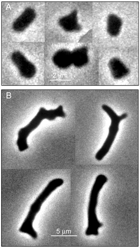
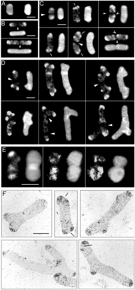
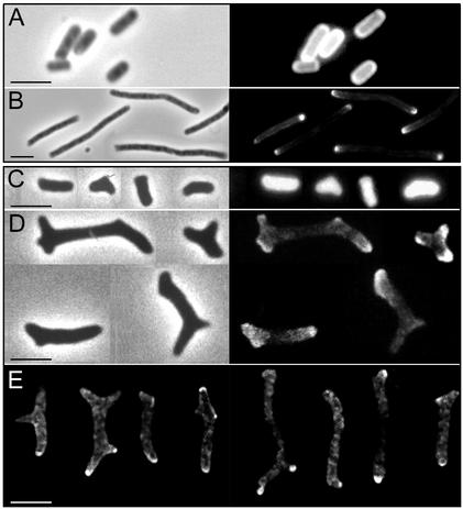
Some strains of Escherichia coli defective for dacA, the gene coding for penicillin-binding protein 5, exhibit a strong branching phenotype when cell division is blocked. Since such branch formation implies a differentiation of polar caps at ectopic locations in the cell envelope, we analyzed murein segregation and observed a strong correlation between areas of inert murein and these morphological anomalies. In particular, the tips of branches exhibited the same properties as those described for polar caps of wild-type cells, i.e., the synthesis and turnover of murein were inhibited. Also, the mobility of cell envelope proteins was apparently constrained in areas with morphological defects. Polar regions of branching cells and sacculi had aberrant morphologies with a very high frequency. Of special interest was that areas of inert murein at polar caps were often split by areas of active synthesis, a situation unlike that observed in wild-type cells. These observations suggest that in dacA mutants, branches and other morphological anomalies may arise from split polar caps or by de novo generation of new poles built around inert peptidoglycan patches in the side walls of the cell.


Figures

FIG. 1.
FIG. 1.
Morphology of E. coli CS801-4 cells in the presence and absence of the cell division inhibitor aztreonam. E. coli CS801-4 cells were grown in LB medium and in LB plus 1 μg of aztreonam per ml for two and a half mass doubling times, and samples were observed and photographed under the phase-contrast microscope. The negatives were digitized, and mosaics with the images of selected cells were constructed with Adobe Photoshop 7.0 (Adobe Systems Inc., San Jose, Calif.) software. (A) Cells from the untreated culture. (B) Cells from the aztreonam-treated culture.
FIG. 2.
FIG. 2.
Segregation of murein in d-cysteine-labeled sacculi from E. coli CS801-4. Cells labeled with d-cysteine were transferred to d-cysteine-free LB medium with and without 1 μg of aztreonam per ml. Samples were removed when the OD550 increased by three- and fivefold, and murein was purified and processed for immunodetection of d-cysteine-containing and total murein as described in the text. As a control, a culture of the parental strain CS109 was d-cysteine labeled and chased in parallel. Samples were removed at the initiation of the chase period and when the OD increased by threefold. Sacculi were observed either by confocal (A to E) or by electron (F) microscopy. Fluorescence pictures were captured in two channels; one would image the distribution of d-cysteine-containing murein (left or upper image in each frame), and the second would image total murein (right or bottom image in each frame). Mosaics depicting selected sacculi were constructed with Adobe Photoshop software. (A) CS801-4 sacculi at the initiation of the chase period. (B) Sacculi from CS109 chased in the presence of aztreonam for a threefold increase in OD. (C) Sacculi from CS801-4 chased in the presence of aztreonam for a threefold increase in OD. (D) Sacculi from CS801-4 chased in the presence of aztreonam for a fivefold increase in OD. (E) Sacculi from CS801-4 chased in the absence of aztreonam for a threefold increase in OD. (F) Sacculi from CS801-4 chased in the presence of aztreonam for a fivefold increase in OD. Silver grains reveal areas of d-cysteine-containing murein. The electron microscopic negatives were digitized and further processed for the mosaic picture with Adobe Photoshop software. Bars, 2.5 μm. Triangular arrowheads indicate potential division sites made up of “all new” murein, V-shaped arrowheads indicate regions of conserved murein outside the poles, and small arrows indicate apparently split poles.
FIG. 3.
FIG. 3.
Segregation of Texas Red succinimidyl ester-labeled surface components. Cells of CS801-4 and its parental strain CS109 were labeled with Texas Red X-succinimidyl ester, transferred to LB medium plus 1 μg of aztreonam per ml, and incubated at 37°C. Samples were removed immediately after dilution and when the OD had increased by fivefold. Cells were fixed and further processed for phase-contrast/epifluorescence or confocal microscopy as described in the text. (A) Unchased cells of CS109; (B) chased cells of CS109; (C) unchased cells of CS801-4; (D) chased cells of CS801-4. Right panels show the fluorescence image for Texas Red in the cells visualized by phase contrast in the left panels. (E) Selected cells of CS801-4 as observed by confocal microscopy. The optical planes showed are those apparently corresponding to the central sections of the cells. It is relevant that CS801-4 cells do not extend flat on the glass slides because they are bent in more than one plane. Nominal thickness of the optical sections was 0.53 μm. Bars, 5 μm. The figure was assembled from digitized photographic slides (phase contrast and epifluorescence) or from digital images (confocal) with Adobe Photoshop software.

Comment in

References

    1. Ayala, J. A., T. Garrido, M. A. de Pedro, and M. Vicente. 1994. Molecular biology of bacterial septation, p. 73-101. In J. M. Ghuysen and R. Hakenbeck (ed.), Bacterial cell wall. Elsevier, Amsterdam, The Netherlands.
    1. Cooper, S. 1991. Bacterial growth and division. Academic Press, Inc., San Diego, Calif.
    1. Danscher, G. 1981. Histochemical demonstration of heavy metals. A revised version of the sulphide silver method suitable for both light and electron microscopy. Histochemistry 71:1-16. - PubMed
    1. Denome, S. A., P. K. Elf, T. A. Henderson, D. E. Nelson, and K. D. Young. 1999. Escherichia coli mutants lacking all possible combinations of eight penicillin binding proteins: Viability, characteristics, and implications for peptidoglycan synthesis. J. Bacteriol. 181:3981-3993. - PMC - PubMed
    1. de Pedro, M. A., J. C. Quintela, J.-V. Höltje, and H. Schwarz. 1997. Murein segregation in Escherichia coli. J. Bacteriol. 179:2823-2834. - PMC - PubMed

Publication types

MeSH terms

LinkOut - more resources