. 2003 Feb;185(4):1174-80.
doi: 10.1128/JB.185.4.1174-1180.2003.

Interactions between phage-shock proteins in Escherichia coli

Affiliations

Interactions between phage-shock proteins in Escherichia coli

Hendrik Adams et al. J Bacteriol. 2003 Feb.

Abstract

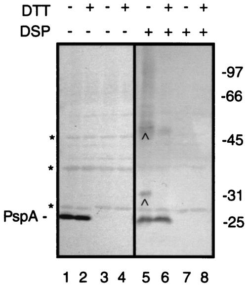
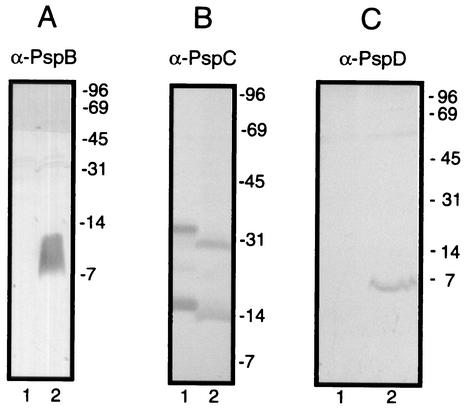
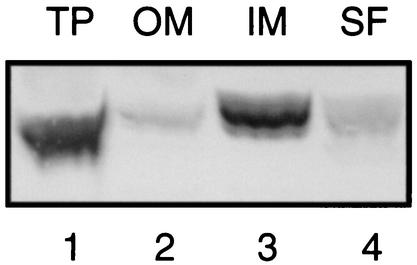
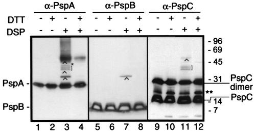
Expression of the pspABCDE operon of Escherichia coli is induced upon infection by filamentous phage and by many other stress conditions, including defects in protein export. Expression of the operon requires the alternative sigma factor sigma54 and the transcriptional activator PspF. In addition, PspA plays a negative regulatory role, and the integral-membrane proteins PspB and PspC play a positive one. In this study, we investigated whether the suggested protein-protein interactions implicated in this complex regulatory network can indeed be demonstrated. Antisera were raised against PspB, PspC, and PspD, which revealed, in Western blotting experiments, that PspC forms stable sodium dodecyl sulfate-resistant dimers and that the hypothetical pspD gene is indeed expressed in vivo. Fractionation experiments showed that PspD localizes as a peripherally bound inner membrane protein. Cross-linking studies with intact cells revealed specific interactions of PspA with PspB and PspC, but not with PspD. Furthermore, affinity-chromatography suggested that PspB could bind PspA only in the presence of PspC. These data indicate that regulation of the psp operon is mediated via protein-protein interactions.


Figures

FIG. 1.
FIG. 1.
Detection of PspA-associated proteins by in vivo cross-linking. Cells of strain CE1224 (lanes 1, 2, 5, and 6) or its pspA::kan derivative CE1343 (lanes 3, 4, 7, and 8), both carrying plasmid pMR05H2, were grown under phosphate limitation to induce the expression of mutant PhoE protein and, consequently, of the psp operon. After incubation of the cells with DSP (lanes 5 to 8), proteins were boiled in sample buffer, with (+) or without (−) DTT as indicated, and separated by SDS-11% PAGE, and this was followed by Western blotting using antibodies directed against PspA. The position of monomeric PspA is indicated. Asterisks depict proteins that nonspecifically react with anti-PspA antiserum. Relevant cross-linked adducts are indicated by carets. Blots were developed with 4-chloro-1-naphthol-H2O2 as the substrate. The positions of molecular mass marker proteins are indicated at the right (in kilodaltons).
FIG. 2.
FIG. 2.
Specificity of anti-PspB, anti-PspC and anti-PspD antisera. (A) Total protein samples of pspB::kan strain CE1417 (lane 1) and strain CE1224 expressing PspB from pJP379 (lane 2) were separated by SDS-20% PAGE, and this was followed by immunoblotting with antiserum directed against PspB (α-PspB) (dilution, 1:1,000). (B) IMVs of strain SG13009[pREP4] expressing His-tagged PspC from pHis-PspC (lane 1) and of strain CE1224 expressing PspABCDE from pJP380 (lane 2) were separated by SDS-15% PAGE, and this was followed by immunoblotting with antiserum directed against PspC (α-PspC) (dilution, 1:1,000). (C) Total protein samples of strain CE1224 carrying pJP119HE (lane 1) or expressing PspABCDE from pJP380 (lane 2) were separated by SDS-15% PAGE, and this was followed by immunoblotting with antiserum directed against PspD (α-PspD) (dilution, 1:1,000). Blots were developed with 4-chloro-1-naphthol-H2O2 as the substrate. The positions of molecular mass marker proteins are indicated at the right of each panel (in kilodaltons).
FIG. 3.
FIG. 3.
Subcellular localization of PspD. Cells of strain CE1224 carrying pJP380 were grown in L-broth, and expression of the plasmid-encoded psp genes was induced by the addition of 100 μM IPTG. After 3 h of incubation, cells were harvested and either directly analyzed (total proteins [TP]) or fractionated. After subjecting the cells to a French press, the outer membrane (OM) and inner membrane (IM) fractions and the soluble fraction (SF) (i.e., the combined periplasmic and cytoplasmic fraction) were obtained by differential ultracentrifugation. The fractions obtained were analyzed by SDS-PAGE followed by Western blotting with anti-PspD antiserum. The blot was developed with 4-chloro-1-naphthol-H2O2 as the substrate.
FIG. 4.
FIG. 4.
Complex formation of Psp proteins. Cells of strain CE1224, expressing PspABCDE from plasmid pJP380, were treated with DSP followed by SDS-PAGE and Western blotting with anti-PspA (α-PspA), anti-PspB (α-PspB), and anti-PspC (α-PspC) antisera. Where indicated, samples were boiled in the presence of DTT prior to SDS-PAGE to reverse the cross-linking. The positions of monomeric PspA, PspB, and PspC and the dimeric form of PspC are indicated. Relevant cross-linked complexes are indicated by carets, square brackets, and a single asterisk. The double asterisk indicates a band that reacts specifically with anti-PspC antiserum, but the exact composition is unknown. The positions of the molecular mass marker proteins are indicated at the right (in kilodaltons). Blots were developed with SuperSignal West Pico chemiluminescent substrate.
FIG. 5.
FIG. 5.
Truncated GST-PspA fusion complements pspA mutant CE1343 for growth on MacConkey plates. Cells of wild-type (wt) strain CE1224 and strain CE1343 (pspA), carrying either vector plasmid pRP269 or pGST-PspAΔC, were streaked on L-broth (A) and MacConkey (B) plates containing ampicillin. The plates were incubated at 30°C for 24 h, and the results were photographed.
FIG. 6.
FIG. 6.
GST-PspA and GST-PspB pull-down assays. Purified GST, GST-PspAΔC, or GST-PspBcyt was bound on a GST-affinity resin, and CHAPS-solubilized IMVs from cells of strain CE1224, overexpressing either PspABCDE, PspA, or PspB from pJP380, pJP381, and pJP379, respectively, were added. After extensive washing, the resin was eluted with glutathione, and eluted proteins were analyzed by SDS-PAGE and either Western blotting with antisera directed against PspA (A) or PspB (B) or silver staining (C). Immunoblots were developed with SuperSignal West Pico chemiluminescent substrate. The positions of GST-PspAΔC, GST-PspBcyt, PspA, PspB, and PspC are indicated. Note that the extract of the PspABCDE overproducer contained equal amounts of PspA and PspB, as did the extracts of the PspA overproducer and of the PspB overproducer, respectively, as determined by Western blotting (data not shown).

Similar articles

Cited by

References

    1. Adams, H., W. Teertstra, M. Koster, and J. Tommassen. 2002. PspE (phage-shock protein E) of Escherichia coli is a rhodanese. FEBS Lett. 518:173-176. - PubMed
    1. Agterberg, M., H. Adriaanse, A. van Bruggen, M. Karperien, and J. Tommassen. 1990. Outer-membrane PhoE protein of Escherichia coli K-12 as an exposure vector: possibilities and limitations. Gene 88:37-45. - PubMed
    1. Akrim, M., M. Bally, G. Ball, J. Tommassen, H. Teerink, A. Filloux, and A. Lazdunski. 1993. Xcp-mediated protein secretion in Pseudomonas aeruginosa: identification of two additional genes and evidence for regulation of xcp gene expression. Mol. Microbiol. 10:431-443. - PubMed
    1. Ansorge, W. 1985. Fast and sensitive detection of protein and DNA bands by treatment with potassium permanganate. J. Biochem. Biophys. Methods 11:13-20. - PubMed
    1. Bauer, M. F., C. Sirrenberg, W. Neupert, and M. Brunner. 1996. Role of Tim23 as voltage sensor and presequence receptor in protein import into mitochondria. Cell 87:33-41. - PubMed

Publication types

MeSH terms

LinkOut - more resources