. 2003 Feb;185(4):1195-207.
doi: 10.1128/JB.185.4.1195-1207.2003.

Analysis of residues determining specificity of Vibrio cholerae TonB1 for its receptors

Affiliations

Analysis of residues determining specificity of Vibrio cholerae TonB1 for its receptors

Alexandra R Mey et al. J Bacteriol. 2003 Feb.

Abstract

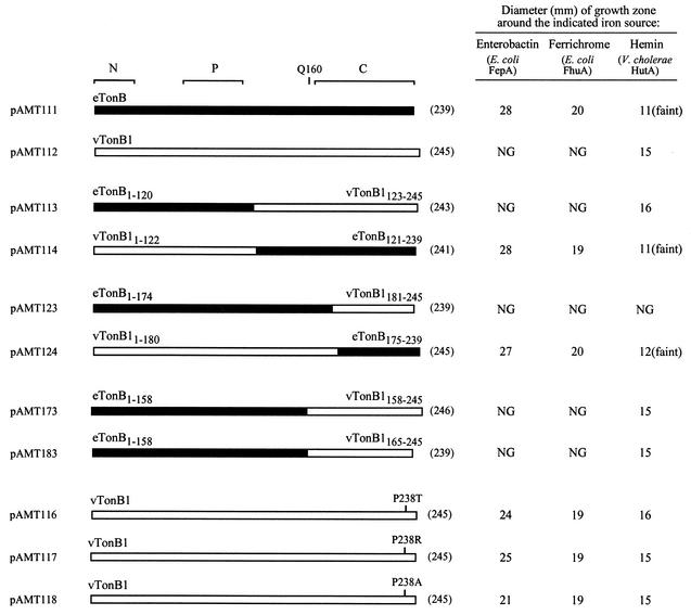
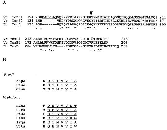
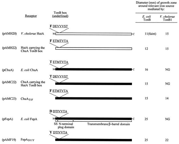
In gram-negative organisms, high-affinity transport of iron substrates requires energy transduction to specific outer membrane receptors by the TonB-ExbB-ExbD complex. Vibrio cholerae encodes two TonB proteins, one of which, TonB1, recognizes only a subset of V. cholerae TonB-dependent receptors and does not facilitate transport through Escherichia coli receptors. To investigate the receptor specificity exhibited by V. cholerae TonB1, chimeras were created between V. cholerae TonB1 and E. coli TonB. The activities of the chimeric TonB proteins in iron utilization assays demonstrated that the C-terminal one-third of either TonB confers the receptor specificities associated with the full-length TonB. Single-amino-acid substitutions near the C terminus of V. cholerae TonB1 were identified that allowed TonB1 to recognize E. coli receptors and at least one V. cholerae TonB2-dependent receptor. This indicates that the very C-terminal end of V. cholerae TonB1 determines receptor specificity. The regions of the TonB-dependent receptors involved in specificity for a particular TonB protein were investigated in experiments involving domain switching between V. cholerae and E. coli receptors exhibiting different TonB specificities. Switching the conserved TonB box heptapeptides at the N termini of these receptors did not alter their TonB specificities. However, replacing the amino acid immediately preceding the TonB box in E. coli receptors with an aromatic residue allowed these receptors to use V. cholerae TonB1. Further, site-directed mutagenesis of the TonB box -1 residue in a V. cholerae TonB2-dependent receptor demonstrated that a large hydrophobic amino acid in this position promotes recognition of V. cholerae TonB1. These data suggest that the TonB box -1 position controls productive interactions with V. cholerae TonB1.


Figures

FIG. 1.
FIG. 1.
Organization of the tonB1 locus in V. cholerae.
FIG. 2.
FIG. 2.
The specificity of TonB for outer membrane receptors is determined by the C terminus of TonB. E. coli TonB (eTonB) is represented by a solid bar; V. cholerae TonB1 (vTonB1) is represented by an open bar. The brackets above the eTonB bar delineate the three functional domains of E. coli TonB: the N-terminal domain (N), the central proline-rich region (P), and the C-terminal dimerization domain (C). Specific residues are shown above the relevant bar in the one-letter amino acid code. The length (in amino acid residues) of each protein is indicated in parentheses on the right. The receptor involved in the utilization of each iron source tested is listed in parentheses below the iron source. Heme utilization was tested in strains supplied with V. cholerae hutA on plasmid pAMH20. The plasmid encoding each TonB protein tested in the assay is listed on the left. E. coli ARM100 or ARM100/pAMH20 carrying the indicated tonB plasmid was seeded at 106 cells per ml into molten L agar containing 100 μg of EDDA per ml. Iron sources were spotted onto the solidified agar as follows: 20 μl of 10 mM FeSO4, 5 μl of 50 μM ferrichrome, 5 μl of DH5α overnight culture (enterobactin), and 5 μl of 50 μM hemin. Plates were incubated for 24 h at 37°C, and the diameter of each zone of growth was measured. Comparable growth around FeSO4 was observed for all strains (data not shown). NG, no growth. “Faint” indicates that this growth zone was less dense than the zones produced by other strains around the specified iron source. The assays were repeated at least three times. Although the relative differences between strains were consistent between experiments, the absolute values for each strain varied due to subtle changes in the medium and assay conditions, and thus only the results of one representative experiment are shown.
FIG. 3.
FIG. 3.
(A) Amino acid sequence alignment of the C-terminal region from V. cholerae TonB1 (Vc TonB1) with the V. cholerae TonB2 (Vc TonB2) and E. coli TonB (Ec TonB) C termini. Identical residues are indicated by an asterisk, and conserved residues are indicated by a dot below the alignment. The numbers flanking the sequences refer to the first and last amino acids in the sequence. The numbering used for the Vc TonB1 sequence is based on the tonB1X sequence, which encodes an extra amino acid compared with the native V. cholerae TonB1 protein (see Materials and Methods). This insertion does not affect the alignment of the sequences shown. The V. cholerae TonB1 region included in the chimera encoded by pAMT173 but not in the chimera encoded by pAMT183 is underlined. The arrowhead marks the site of the C-terminal domain switch in the chimeras encoded by pAMT123 and pAMT124. The V. cholerae TonB1 Pro238 residue is shown in boldface. (B) Comparison of the predicted TonB box regions from E. coli and V. cholerae receptors. The TonB box heptapeptide is underlined. The TonB box −1 residue is shown in boldface.
FIG. 4.
FIG. 4.
Analysis of receptor residues necessary for productive interactions with E. coli TonB or V. cholerae TonB1. Solid bars represent E. coli receptors; open bars represent V. cholerae receptors. The domain organization shown for FepA (signal sequence [SS], N-terminal plug domain, and β-barrel domain) is expected to be relevant for all the receptors shown. The TonB box sequence (underlined), as well as the residue immediately preceding the TonB box, is shown above each bar. The plasmid encoding each receptor protein is listed on the left. The numbering scheme used to designate the TonB box −1 position in FepA and ChuA reflects the amino acid position in the mature (cleaved) protein. E. coli ARM100 carrying the indicated receptor plasmid, and a plasmid encoding either E. coli TonB or V. cholerae TonB1, was tested as described in the legend to Fig. 2. The function of the HutA or ChuA receptors was tested with hemin as the iron source. The function of the FepA receptors was tested with enterobactin as the iron source. Comparable growth around FeSO4 was observed for all strains (data not shown). NG, no growth. “Faint” indicates that this growth zone was less dense than the zones produced by other strains around the specified iron source. The assays were repeated at least three times. Although the relative differences between strains were consistent between experiments, the absolute values for each strain varied due to subtle changes in the medium and assay conditions, and thus only results from one representative experiment are shown.
FIG. 5.
FIG. 5.
Expression of wild-type (wt) IrgA and IrgA point mutants in V. cholerae. Samples representing 5 × 10⁶ cells were prepared as described in Materials and Methods and resolved by SDS-7.5% PAGE. Immunoblotting was performed with polyclonal anti-IrgA antiserum. Lane 1, CA40130N; lane 2, ARM616/pWKS30; lane 3, ARM616/pCAT121; lane 4, ARM616/pAMI2; lane 5, ARM616/pAMI3; lane 6, ARM616/pAMI4.

Similar articles

Cited by

References

    1. Barnard, T. J., M. E. Watson, and M. A. McIntosh. 2001. Mutations in the Escherichia coli receptor FepA reveal residues involved in ligand binding and transport. Mol. Microbiol. 41:527-536. - PubMed
    1. Bell, P. E., C. D. Nau, J. T. Brown, J. Konisky, and R. J. Kadner. 1990. Genetic suppression demonstrates interaction of TonB protein with outer membrane transport proteins in Escherichia coli. J. Bacteriol. 172:3826-3829. - PMC - PubMed
    1. Bradbeer, C. 1993. The proton motive force drives the outer membrane transport of cobalamin in Escherichia coli. J. Bacteriol. 175:3146-3150. - PMC - PubMed
    1. Braun, M., H. Killmann, and V. Braun. 1999. The β-barrel domain of FhuΔ5-160 is sufficient for TonB-dependent FhuA activities of Escherichia coli. Mol. Microbiol. 33:1037-1049. - PubMed
    1. Braun, V. 1995. Energy-coupled transport and signal transduction through the gram-negative outer membrane via TonB-ExbB-ExbD-dependent receptor proteins. FEMS Microbiol. Rev. 16:295-307. - PubMed

Publication types

MeSH terms

LinkOut - more resources