. 2003 Feb;185(4):1273-83.
doi: 10.1128/JB.185.4.1273-1283.2003.

Transcriptional switch on of ssgA by A-factor, which is essential for spore septum formation in Streptomyces griseus

Affiliations

Transcriptional switch on of ssgA by A-factor, which is essential for spore septum formation in Streptomyces griseus

Haruka Yamazaki et al. J Bacteriol. 2003 Feb.

Abstract

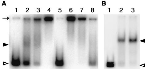
A-factor (2-isocapryloyl-3R-hydroxymethyl-gamma-butyrolactone) triggers morphological development and secondary metabolism in Streptomyces griseus. A transcriptional activator (AdpA) in the A-factor regulatory cascade switches on a number of genes required for both processes. AdBS11 was identified in a library of the DNA fragments that are bound by AdpA and mapped upstream of ssgA, which is essential for septum formation in aerial hyphae. Gel mobility shift assays and DNase I footprinting revealed three AdpA-binding sites at nucleotide positions about -235 (site 1), -110 (site 2), and +60 (site 3) with respect to the transcriptional start point, p1, of ssgA. ssgA had two transcriptional start points, one starting at 124 nucleotides (p1) and the other starting at 79 nucleotides (p2) upstream of the start codon of ssgA. Of the three binding sites, only sites 1 and 2 were required for transcriptional activation of p1 and p2 by AdpA. The transcriptional switch on of ssgA required the extracytoplasmic function sigma factor, sigma(AdsA), in addition to AdpA. However, it was unlikely that sigma(AdsA) recognized the two ssgA promoters, since their -35 and -10 sequences were not similar to the promoter sequence motifs recognized by sigma(BldN), a sigma(AdsA) homologue of Streptomyces coelicolor A3(2). An ssgA disruptant formed aerial hyphae, but did not form spores, irrespective of the carbon source of the medium, which indicated that ssgA is a member of the whi genes. Transcriptional analysis of ssfR, located just upstream of ssgA and encoding an IclR-type transcriptional regulator, suggested that no read-through from ssfR into ssgA occurred, and ssgA was transcribed in the absence of ssfR. ssgA was thus found to be controlled by AdpA and not by SsfR to a detectable extent. SsfR appeared to regulate spore septum formation independently of SsgA or through interaction with SsgA in some unknown way, because an ssfR disruptant also showed a whi phenotype.


Figures

FIG. 1.
FIG. 1.
Binding of AdpA-H to AdBS11 (A) and to a region downstream of AdBS11 (B). (A) AdBS11 positioned at −306 to +69 (see Fig. 4B) was 32P labeled and used in the gel mobility shift assay. The amounts of AdpA-H used in lanes 1, 2, 3, and 4 were 0, 0.04, 0.2, and 0.4 μg, respectively. Lanes 5 to 8 show competition of binding between AdBS11 and AdpA-H by an excess amount of nonlabeled probe. The amounts of AdpA-H used were 0 μg (lane 5) and 0.4 μg (lanes 6, 7, and 8). The concentrations of nonlabeled probes added to lanes 7 and 8 were 100× and 500×, respectively. The positions of free probes (open arrowhead), AdpA-H-bound probes (solid arrowhead), and the gel well (arrow) are shown. (B) Probe dw at positions −25 to +198 (Fig. 4B) was 32P labeled and used in the gel mobility shift assay. The amounts of AdpA-H used in lanes 1, 2, and 3 were 0, 0.2, and 0.8 μg, respectively.
FIG. 2.
FIG. 2.
ORFs in the cloned 4.6-kb SalI fragment (A), nucleotide sequence of the region covering the ssgA promoter and a 3′ portion of ssfR (B), and low-resolution S1 nuclease mapping of ssgA in S. griseus (C and D). (A) The positions and directions of ORFs predicted by Frame Plot analysis (12) of the nucleotide sequence are indicated by arrows. The position of AdBS11 is also shown. (B) The nucleotide number is shown, taking the transcriptional start point of ssgA p1 as +1. The amino acid sequences of a C-terminal region of SsfR and an N-terminal region of SsgA are also shown. The AdBS11 region (nucleotide positions −306 to +69) is indicated by arrows. The two transcriptional start points p1 and p2 are shown in lowercase letters. Three AdpA-H-binding sites (Fig. 4) are indicated by lines. The upper lines indicate the sense strands protected from DNase I digestion, and the lower lines indicate the antisense strand protected. Three SacI sites described in the legend to Fig. 6C are boxed. (C) RNA was prepared from cells grown at 28°C for the indicated number of days on YMPD agar medium in the wild-type S. griseus IFO13350 (wt), an adpA disruptant (ΔadpA), an A-factor-deficient mutant strain (HH1), and an adsA disruptant (ΔadsA). hrdB, which is transcribed at rather constant levels throughout growth, served as an internal control. ssgA was transcribed from two sites (p1 and p2) in the wild type and at a very low level in the ΔadpA, HH1, and ΔadsA strains. (D) Transcription of ssgA, as well as adpA, is enhanced in the wild-type strain harboring pADP10H.
FIG. 3.
FIG. 3.
Transcriptional start point of ssgA. (A) Two transcriptional start points of ssgA, as determined by high-resolution S1 mapping. RNA prepared from the wild-type cells grown at 28°C for 3 days on YMPD agar medium was used. The arrowheads indicate the positions of the S1-protected fragments. The 5′ termini of the transcripts were assigned to the T (+1) and A (+46) residues indicated in lowercase letters, because the fragments generated by the chemical sequencing reactions migrate 1.5 nucleotides further than the corresponding fragments generated by S1 nuclease digestion of the DNA-RNA hybrids (half a residue from the presence of the 3′-terminal phosphate group and one residue from the elimination of the 3′-terminal nucleotide). (B) Transcriptional analysis of ssgA by RT-PCR. RNA was prepared from the wild-type cells grown at 30°C for 26 h in YMPD liquid medium or at 28°C for 2 days on YMPD agar medium. After RT-PCR with 25 cycles of amplification, the amplified fragments were separated by 2% agarose gel electrophoresis. RT-PCR was also performed similarly in the absence of reverse transcriptase, as indicated by a minus sign. Probe 2 was used for detection of the transcript from p1, and probe 3 was used for detection of the transcripts from p1 and p2. Transcriptional read-through from ssfR was not detectable with probe 1. (C) The promoter sequence of bldM p1, regulated by σBldN (4), a σAdsA homologue in S. coelicolor A3(2), is aligned with those of ssgA p1 and p2. The predicted −10 and −35 sites in the promoters are underlined. The identical nucleotide residues between ssgA p1 and p2 are indicated by asterisks. Transcriptional start points are also underlined.
FIG. 4.
FIG. 4.
Three AdpA-binding sites located upstream of ssgA, as determined by DNase I footprinting. (A) DNase I footprinting assays were performed on the sense strand (+) and the antisense strand (−) probes. The amounts of AdpA-H used in lanes 1, 2, 3, 4, and 5 were 0, 0.3, 0.6, 0.9, and 0 μg for sites 1 and 2 and 0, 0.3, 0.4, 0.6, and 0 μg for site 3. (B) The positions of the AdpA-binding sites relative to the ssgA and ssfR coding sequences are shown. Sites 1, 2, and 3 center at positions −235, −110, and +60, respectively. AdBS11 and dw were used as the probes for the gel mobility shift assay in Fig. 1.
FIG. 5.
FIG. 5.
Mutational analysis of the AdpA-binding sites. (A) Mutations introduced in the AdpA-binding sites. An XhoI site generated in sites 1 and 2 and a BamHI site generated in site 3 are indicated by italic letters. m1, m2, and m3 represent the mutations introduced in the AdpA-binding sites 1, 2, and 3, respectively. (B) Probes pb11, -22, -33, -12, and -23 were prepared by PCR with pKF1 to pKF7 as the templates and used as the 32P-labeled probes for the gel mobility shift assays in panel C. (C) Gel mobility shift assays for determination of AdpA binding to mutated sequences. (Top) The amounts of AdpA-H used in lanes 1, 2, 3, 4, and 5 were 0, 60, 150, 300, and 900 ng, respectively. AdpA-H bound pb11 (containing the intact AdpA-binding site) prepared by PCR with pKF1, whereas it failed to bind pb11 containing mutated sequence m1 similarly prepared with pKF2. Similarly, AdpA-H did not bind mutated sequence m2 or m3. The positions of free (open arrowhead) and AdpA-H-bound (solid arrowhead) probes and the gel well (arrow) are shown. (Middle) Gel mobility shift assays with pb12. The amounts of AdpA-H used in lanes 1, 2, 3, 4, and 5 were 0, 15, 45, 150, and 300 ng, respectively. The templates used for PCR for preparing pb12 are indicated above the panel. When pb12 was prepared with pKF1 as the template, it contained two (sites 1 and 2) of the three AdpA-binding sites and gave two retarded signals. (Bottom) Gel mobility shift assays with pb23. The amounts of AdpA-H were the same as those in the middle panel. The templates used are indicated above the panel.
FIG. 6.
FIG. 6.
Requirement of both sites 1 and 2 for the transcriptional activation of ssgA by AdpA. (A) Construction of mutant ΔssgA. The restriction sites of SmaI, PstI, and SalI are abbreviated as Sm, Pt, and Sl, respectively. The DNA fragment on pKgA used for complementation analysis is also shown. (B) Southern hybridization to check correct disruption with 32P-labeled probes 1 and 3 against the SmaI-digested chromosomal DNA from the wild type (wt) and the ΔssgA mutant. (C) Schematic representation of low-resolution S1 mapping of the ssgA promoters on plasmids in the ΔssgA background. The SacI and PstI sites are indicated as Sc and Pt, respectively. One of the three SacI sites (GAGCTC) was mutated to GAGCTG for experimental convenience in construction of the pKGA-series plasmids. This mutation does not change the amino acid. (D) Transcription of ssgA in the ΔssgA mutants harboring pKGA1 to pKGA8. RNA was prepared from cells grown on YMPD agar medium at 28°C for the indicated days. Two transcripts with the same sizes as those detected in the wild type were detected only in the ΔssgA strain harboring pKGA1 and pKGA4.
FIG. 7.
FIG. 7.
Scanning electron micrographs of ΔssgA transformants. wt, wild type. Strains were grown on R2YE agar medium at 28°C for 3 days. The defect in spore formation in the ΔssgA strains was complemented by pKGA1 and pKGA4, but not by pKGA2. Bar, 2 μm.
FIG. 8.
FIG. 8.
Transcription of ssgA in the absence of ssfR. (A) Construction of the mutant ΔssfR strain. The restriction sites of SmaI, SalI, PmaCI, and EcoT14I are abbreviated Sm, Sl, Pm, and EcT, respectively. The DNA fragment on pKfR used for complementation analysis is also shown. (B) Southern hybridization with 32P-labeled probes 2 and 3 against the SmaI-digested chromosomal DNA from the wild type and ΔssfR strain to confirm correct gene replacement by homologous recombination. (C) Scanning electron micrographs of S. griseus ΔssfR strains with and without pKfR. Bar, 1 μm. Strains were grown on YMPD agar medium at 28°C for 3 days. The defect in spore formation of ΔssfR mutants is complemented by a low-copy-number plasmid, pKfR, containing the whole ssfR sequence. (D) Transcription of ssgA in the wild-type strain grown at 28°C for the indicated days on YMPD or YMP-mannitol medium and in the ΔssfR mutant grown on YMPD medium.

Similar articles

Cited by

References

    1. Ando, N., K. Ueda, and S. Horinouchi. 1997. A Streptomyces griseus gene (sgaA) suppresses the growth disturbance caused by high osmolality and a high concentration of A-factor during early growth. Microbiology 143:2715-2723. - PubMed
    1. Ausubel, F. M., R. Brent, R. E. Kingston, D. O. Moore, J. S. Seidman, J. A. Smith, and K. Struhl. 1987. Current protocols in molecular biology. John Wiley & Sons, Inc., New York, N.Y.
    1. Beck, E., G. Ludwig, E. A. Auerswald, B. Reiss, and H. Schaller. 1982. Nucleotide sequence and exact localization of the neomycin phosphotransferase gene from transposon Tn5. Gene 19:327-336. - PubMed
    1. Bibb, M. J., V. Molle, and M. J. Buttner. 2000. σBldN, an extracytoplasmic function RNA polymerase sigma factor required for aerial mycelium formation in Streptomyces coelicolor A3(2). J. Bacteriol. 182:4606-4616. - PMC - PubMed
    1. Gallegos, M. T., R. Schleif, A. Bairoch, K. Hofmann, and J. L. Ramos. 1997. AraC/XylS family of transcriptional regulators. Microbiol. Mol. Biol. Rev. 61:393-410. - PMC - PubMed

Publication types

MeSH terms

Associated data

LinkOut - more resources