. 2003 Feb;185(4):1423-31.
doi: 10.1128/JB.185.4.1423-1431.2003.

Peptidoglycan synthesis in the absence of class A penicillin-binding proteins in Bacillus subtilis

Affiliations

Peptidoglycan synthesis in the absence of class A penicillin-binding proteins in Bacillus subtilis

Derrell C McPherson et al. J Bacteriol. 2003 Feb.

Abstract

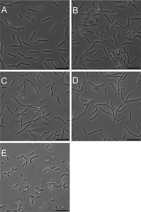
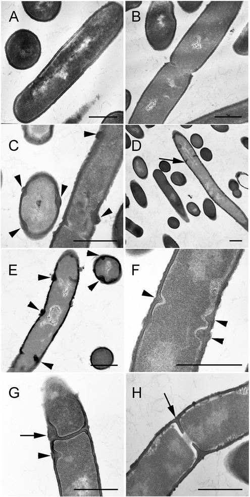
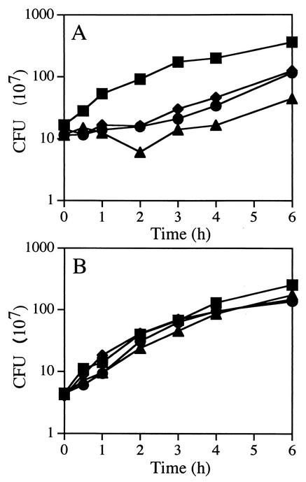
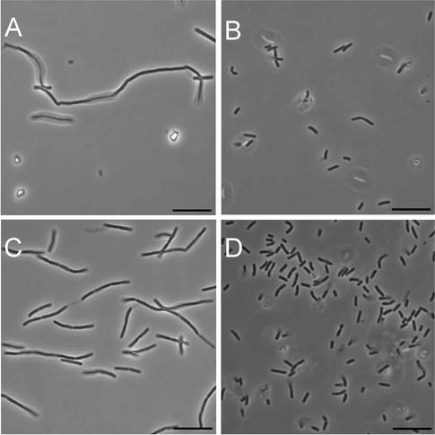
Penicillin-binding proteins (PBPs) catalyze the final, essential reactions of peptidoglycan synthesis. Three classes of PBPs catalyze either trans-, endo-, or carboxypeptidase activities on the peptidoglycan peptide side chains. Only the class A high-molecular-weight PBPs have clearly demonstrated glycosyltransferase activities that polymerize the glycan strands, and in some species these proteins have been shown to be essential. The Bacillus subtilis genome sequence contains four genes encoding class A PBPs and no other genes with similarity to their glycosyltransferase domain. A strain lacking all four class A PBPs has been constructed and produces a peptidoglycan wall with only small structural differences from that of the wild type. The growth rate of the quadruple mutant is much lower than those of strains lacking only three of the class A PBPs, and increases in cell length and frequencies of wall abnormalities were noticeable. The viability and wall production of the quadruple-mutant strain indicate that a novel enzyme can perform the glycosyltransferase activity required for peptidoglycan synthesis. This activity was demonstrated in vitro and shown to be sensitive to the glycosyltransferase inhibitor moenomycin. In contrast, the quadruple-mutant strain was resistant to moenomycin in vivo. Exposure of the wild-type strain to moenomycin resulted in production of a phenotype similar to that of the quadruple mutant.


Figures

FIG. 1.
FIG. 1.
Phase-contrast microscopy of class A PBP mutants. Cells were photographed from exponentially growing (A to D) and 24-h (E) cultures of PS832 (wild type [A]), PS2062 (PBP 1 [B]), DPVB69 (PBP 1 PBP 2c PBP 4 [C]) and DPVB87 (PBP 1 PBP 2c PBP 2d PBP 4 [D and E]). Bars, 10 μm.
FIG. 2.
FIG. 2.
Cell length distributions of class A PBP mutants. Cell were harvested from exponentially growing cultures and examined under phase-contrast microscopy. Cell lengths (n = 200 for each strain) were determined from digital images. (A) PS832 (wild type); (B) PS2062 (PBP 1); (C) DPVB69 (PBP 1 PBP 2c PBP 4); (D) DPVB87 (PBP 1 PBP 2c PBP 2d PBP 4). In the wild-type culture 3% of the cells were ≥10 μm. This increased to 9% in the PBP 1 culture, 20% in the PBP 1 PBP 2c PBP 4 culture, and 33% in PBP 1 PBP 2c PBP 2d PBP 4 cultures.
FIG. 3.
FIG. 3.
Electron microscopy of class A PBP mutants. Cells obtained from exponentially growing cultures of PS832 (wild type [A and B]), PS2062 (PBP 1 [C and D]), and DPVB87 (PBP 1 PBP 2c PBP 2d PBP 4 [E to H]) were fixed, sectioned, and stained for transmission electron microscopy. Arrowheads in C, E, F, and G indicate abnormal cell wall material. Arrows in G indicate irregularly formed septa, and in D and H they indicate septa impinging upon the chromosome. Bars, 0.5 μm.
FIG. 4.
FIG. 4.
Effects of moenomycin on growth of B. subtilis. Cultures of PS832 (wild type [A]) and DPVB87 (PBP 1 PBP 2c PBP 2d PBP 4 [B]) were exposed to either 0 (▪), 16 (•), 40 (♦), or 80 (▴) μg of moenomycin/ml. CFU values are averages from three separate experiments. Moenomycin was added when the OD reached 0.5. At this culture density, growth of the wild-type strain had begun to slow, reducing the difference between the growth rates of the wild-type and quadruple-mutant strains.
FIG. 5.
FIG. 5.
Effects of moenomycin on the morphology of B. subtilis. PS832 (wild type [A and B]) and DPVB87 (PBP 1 PBP 2c PBP 2d PBP 4 [C and D]) cells were exposed to 80 μg of moenomycin/ml for 2.5 h (A and C) and 24 h (B and D) and examined by phase-contrast microscopy. Bars, 10 μm.

References

    1. Abhayawardhane, Y., and G. C. Stewart. 1995. Bacillus subtilis possesses a second determinant with extensive sequence similarity to the Escherichia coli mreB morphogene. J. Bacteriol. 177:765-773. - PMC - PubMed
    1. Adam, M., C. Fraipont, N. Rhazi, M. Nguyen-Disteche, B. Lakaye, J. M. Frere, B. Devreese, J. Van Beeumen, Y. van Heijenoort, J. van Heijenoort, and J. M. Ghuysen. 1997. The bimodular G57-V577 polypeptide chain of the class B penicillin-binding protein 3 of Escherichia coli catalyzes peptide bond formation from thiolesters and does not catalyze glycan chain polymerization from the lipid II intermediate. J. Bacteriol. 179:6005-6009. - PMC - PubMed
    1. Anagnostopoulos, C., and J. Spizizen. 1961. Requirements for transformation in Bacillus subtilis. J. Bacteriol. 81:74-76. - PMC - PubMed
    1. Archibald, A. R., I. C. Hancock, and C. R. Harwood. 1993. Cell wall structure, synthesis, and turnover, p. 381-410. In A. L. Sonenshein, J. A. Hoch, and R. Losick (ed.), Bacillus subtilis and other gram-positive bacteria. American Society for Microbiology, Washington, D.C.
    1. Atrih, A., G. Bacher, G. Allmaier, M. P. Williamson, and S. J. Foster. 1999. Analysis of peptidoglycan structure from vegetative cells of Bacillus subtilis 168 and role of PBP 5 in peptidoglycan maturation. J. Bacteriol. 181:3956-3966. - PMC - PubMed

Publication types

MeSH terms

LinkOut - more resources