

Proteomic analysis of the spore coats of Bacillus subtilis and Bacillus anthracis
- PMID: 12562816
- PMCID: PMC142864
- DOI: 10.1128/JB.185.4.1443-1454.2003
Proteomic analysis of the spore coats of Bacillus subtilis and Bacillus anthracis
Abstract
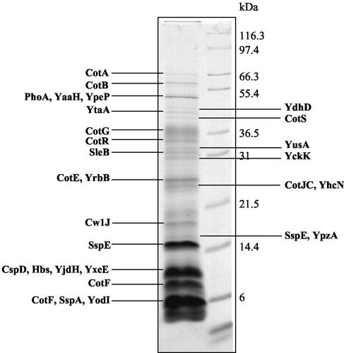
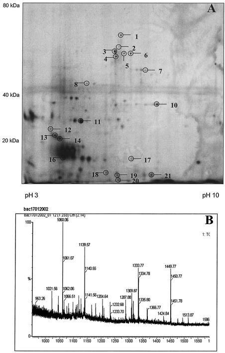
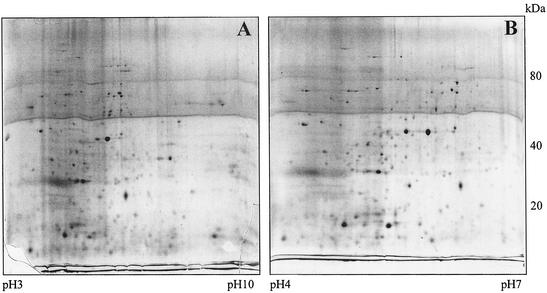
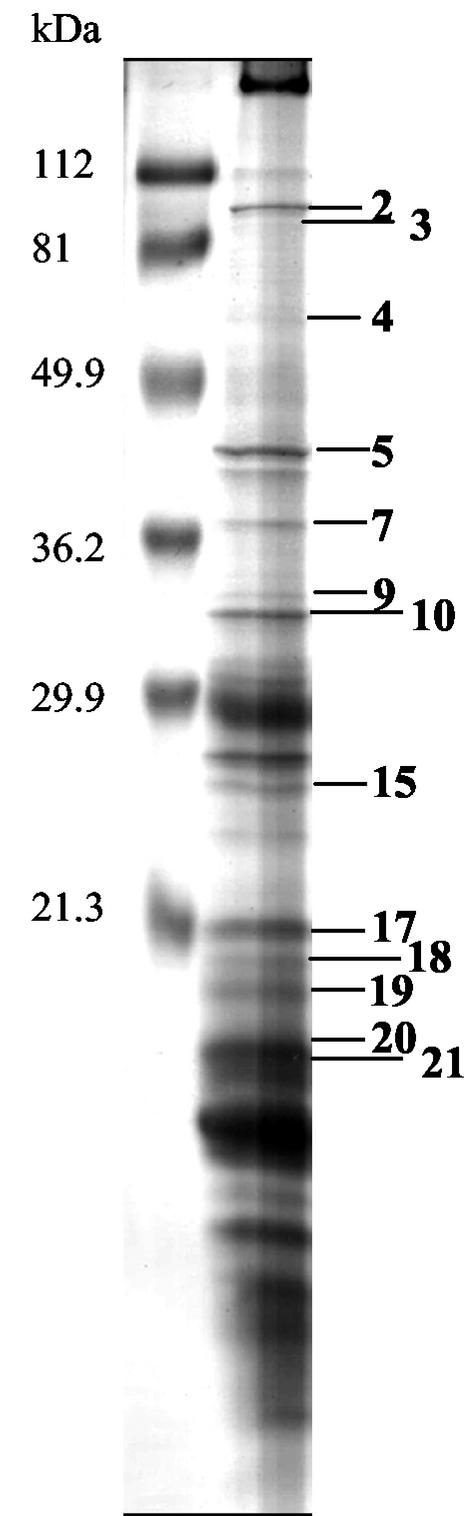
The outermost proteinaceous layer of bacterial spores, called the coat, is critical for spore survival, germination, and, for pathogenic spores, disease. To identify novel spore coat proteins, we have carried out a preliminary proteomic analysis of Bacillus subtilis and Bacillus anthracis spores, using a combination of standard sodium dodecyl sulfate-polyacrylamide gel electrophoresis separation and improved two-dimensional electrophoretic separations, followed by matrix-assisted laser desorption ionization-time of flight and/or dual mass spectrometry. We identified 38 B. subtilis spore proteins, 12 of which are known coat proteins. We propose that, of the novel proteins, YtaA, YvdP, and YnzH are bona fide coat proteins, and we have renamed them CotI, CotQ, and CotU, respectively. In addition, we initiated a study of coat proteins in B. anthracis and identified 11 spore proteins, 6 of which are candidate coat or exosporium proteins. We also queried the unfinished B. anthracis genome for potential coat proteins. Our analysis suggests that the B. subtilis and B. anthracis coats have roughly similar numbers of proteins and that a core group of coat protein species is shared between these organisms, including the major morphogenetic proteins. Nonetheless, a significant number of coat proteins are probably unique to each species. These results should accelerate efforts to develop B. anthracis detection methods and understand the ecological role of the coat.
Figures
References
-
- Abe, A., H. Koide, T. Kohno, and K. Watabe. 1995. A Bacillus subtilis spore coat polypeptide gene, cotS. Microbiology 141:1433-1442. - PubMed
-
- Aizawa, S.-I. 2001. Bacterial flagella and type III secretion systems. FEMS Microbiol. Lett. 202:157-164. - PubMed
-
- Aronson, A. I., H. Y. Song, and N. Bourne. 1989. Gene structure and precursor processing of a novel Bacillus subtilis spore coat protein. Mol. Microbiol. 3:437-444. - PubMed
Publication types
MeSH terms
Substances
Grants and funding
LinkOut - more resources
Full Text Sources
Other Literature Sources
Molecular Biology Databases
