Comparative Study
. 2003 Apr 1;548(Pt 1):203-18.
doi: 10.1113/jphysiol.2002.034447. Epub 2003 Jan 31.

Polarized distribution of HCO3- transport in human normal and cystic fibrosis nasal epithelia

Affiliations
Comparative Study

Polarized distribution of HCO3- transport in human normal and cystic fibrosis nasal epithelia

Anthony M Paradiso et al. J Physiol. .

Abstract

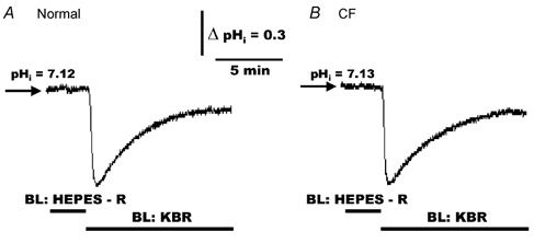
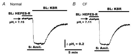
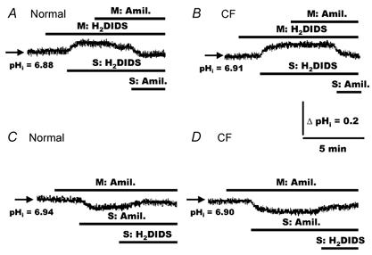
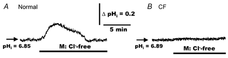
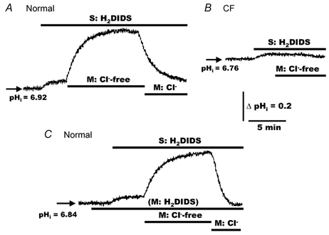
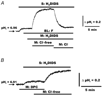
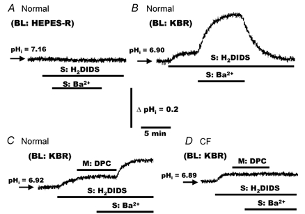
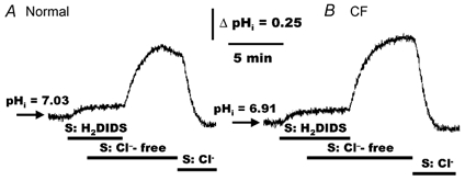
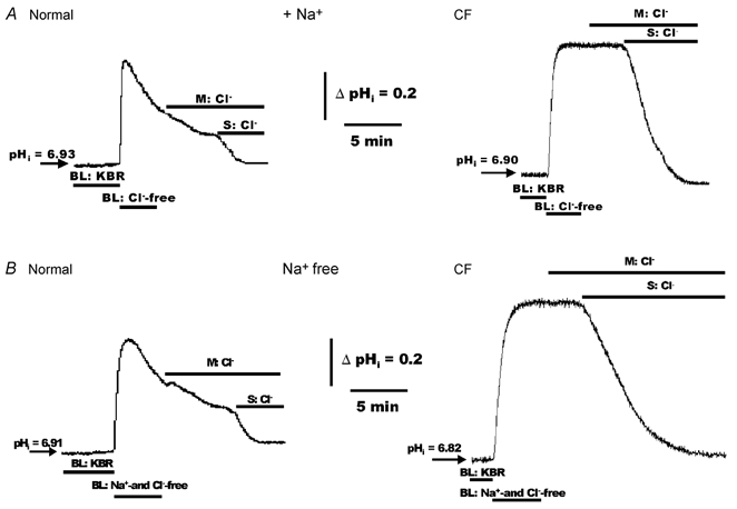
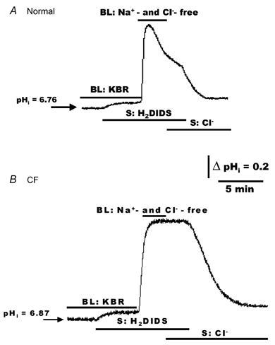
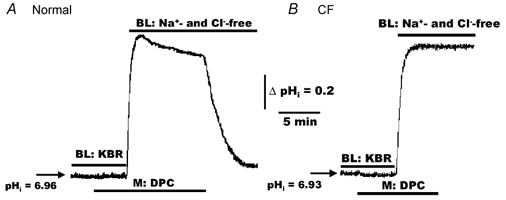
The polarized distribution of HCO3- transport was investigated in human nasal epithelial cells from normal and cystic fibrosis (CF) tissues. To test for HCO3- transport via conductive versus electroneutral Cl-/HCO3- exchange (anion exchange, AE) pathways, nasal cells were loaded with the pH probe 2',7'-bis(carboxyethyl)-5(6)-carboxyfluorescein and mounted in a bilateral perfusion chamber. In normal, but not CF, epithelia, replacing mucosal Cl- with gluconate caused intracellular pH (pHi) to increase, and the initial rates (Delta pH min-1) of this increase were modestly augmented (approximately 26 %) when normal cells were pretreated with forskolin (10 microM). Recovery from this alkaline shift was dependent on mucosal Cl-, was insensitive to the AE inhibitor 4,4'-diisothiocyanatodihydrostilbene-2,2'-disulfonic acid (H2DIDS; 1.5 mM), but was sensitive to the cystic fibrosis transmembrane conductance regulator (CFTR) channel inhibitor diphenylamine-2-carboxylate (DPC; 100 microM). In contrast, removal of serosal Cl- caused pHi to alkalinize in both normal and CF epithelia. Recovery from this alkaline challenge was dependent on serosal Cl- and blocked by H2DIDS. Additional studies showed that serosally applied Ba2+ (5.0 mM) in normal, but not CF, cells induced influx of HCO3- across the apical membrane that was reversibly blocked by mucosal DPC. In a final series of studies, normal and CF cells acutely alkaline loaded by replacing bilateral Krebs bicarbonate Ringer (KBR) with Hepes-buffered Ringer solution exhibited basolateral, but not apical, recovery from an alkaline challenge that was dependent on Cl-, independent of Na+ and blocked by H2DIDS. We conclude that: (1) normal, but not CF, nasal epithelia have a constitutively active DPC-sensitive HCO3- influx/efflux pathway across the apical membrane of cells, consistent with the movement of HCO3- via CFTR; and (2) both normal and CF nasal epithelia have Na+-independent, H2DIDS-sensitive AE at their basolateral domain.


Figures

Figure 1
Figure 1. Basal intracellular pH (pHi) in polarized monolayers of normal and cystic fibrosis (CF) human nasal epithelial (HNE) cells bilaterally perfused with nominally CO2/HCO3-free Ringer solution or Krebs bicarbonate Ringer (KBR)
For both normal (A) and CF (B) cell preparations, HNE cells were initially bilaterally (BL) perfused with Hepes-buffered NaCl Ringer solution (pH 7.4) before changing to bilateral KBR (pH 7.4) at the times indicated in the tracings. Each trace is representative of ten separate experiments (four different individuals).
Figure 2
Figure 2. Effects of amiloride on pHi in response to a CO2/HCO3 challenge in polarized monolayers of normal and CF HNE cells
For both normal (A) and CF (B) cell preparations, HNE cells were initially bilaterally perfused with Hepes-buffered NaCl Ringer solution (pH 7.4) before adding amiloride (Amil., 500 μm) to the serosal (S) perfusate. In the presence of amiloride, the perfusate was subsequently changed to bilateral KBR (pH 7.4) and amiloride removed from the serosal compartment at the times indicated in the tracings. Each trace is representative of seven separate experiments (three different individuals).
Figure 3
Figure 3. Response of pHi to mucosal and serosal additions of H2DIDS and amiloride in polarized monolayers of normal and CF HNE cells
For both normal (A and C) and CF (B and D) cell preparations, HNE cells were bilaterally perfused with KBR before adding 4,4′-diisothiocyanatodihydrostilbene-2,2′-disulfonic acid (H2DIDS; 1.5 mm) and amiloride (500 μm) to the mucosal (M) and serosal (S) compartments as indicated in the tracings. Each trace is representative of seven separate experiments (three different individuals).
Figure 4
Figure 4. Effects of mucosal Cl-free Ringer solution on pHi in polarized monolayers of normal and CF HNE cells
For both normal (A) and CF (B) cell preparations, HNE cells were initially bilaterally perfused with KBR before changing to mucosal (M) Cl-free (gluconate replacing Cl; constant 5 % PCO2/25 mm HCO3, pH 7.4) Ringer solution at the times indicated in the tracings. The serosal perfusate remained KBR throughout these studies. A, representative example of seven separate experiments (four different individuals). B, representative example of eight separate experiments (three different individuals).
Figure 5
Figure 5. Effects of mucosal Cl substitution on pHi in polarized monolayers of normal and CF HNE cells exposed to serosal and/or mucosal H2DIDS
For both normal (A and C) and CF (B) HNE cell preparations, cells were initially perfused bilaterally with KBR and H2DIDS (1.5 mm) added to the serosal and/or mucosal compartment before removing Cl (gluconate replacing Cl; constant 5 % PCO2/25 mm HCO3, pH 7.4) or re-adding Cl to the mucosal perfusate, as shown in the tracings. The serosal perfusate remained KBR throughout the study. A and C, representative examples of eleven separate experiments (four different individuals). B, is representative of eight separate experiments (three different individuals).
Figure 6
Figure 6. Effects of forskolin and diphenylamine-2-carboxylate (DPC) on pHi in response to mucosal Cl substitution in polarized monolayers of normal HNE cells
A, normal HNE cells were initially perfused with bilateral (BL) KBR. Serosal H2DIDS (1.5 mm) and bilateral forskolin (F; 10 μm) were added to the perfusate at the times indicated in the tracing, before changing the mucosal perfusate from KBR to Cl-free (gluconate replacing Cl; constant 5 % PCO2/25 mm HCO3, pH 7.4) KBR. Cl was subsequently re-added to the mucosal compartment at the time indicated in the tracing. B, normal HNE cells were bilaterally perfused with KBR and mucosal DPC (100 μm) and serosal H2DIDS (1.5 mm) added to the perfusate at the times indicated in the tracing. In the presence of DPC, changing the mucosal perfusate from KBR to Cl-free KBR elicited no change in pHi, whereas removal of DPC induced an alkalinization of the cells. For both traces, the serosal perfusate remained KBR throughout the study. A, representative example of seven separate experiments (three different individuals). B, representative example of seven separate experiments (four different individuals).
Figure 7
Figure 7. Effects of mucosal DPC on pHi in response to serosal Ba2+ in polarized monolayers of normal and CF HNE cells
A, normal HNE cells were bilaterally perfused with Hepes-buffered NaCl Ringer solution before adding H2DIDS (1.5 mm) and Ba2+ (5.0 mm) to the serosal compartment at the times shown in the trace. For both normal (B and C) and CF (D) HNE cell preparations, cells were perfused bilaterally with KBR and H2DIDS (1.5 mm) administrated to the serosal perfusate. Following H2DIDS treatment, DPC (100 μm) and/or Ba2+ (5.0 mm) were added to and/or removed from the serosal and/or mucosal compartment in both normal (B and C) and CF (D) cell preparations at the times shown in the tracings. Each trace is representative of six separate experiments (three different individuals).
Figure 8
Figure 8. Effects of H2DIDS and serosal Cl substitution on pHi in polarized monolayers of normal and CF HNE cells
For both normal (A) and CF (B) HNE cell preparations, cells were initially perfused bilaterally with KBR and H2DIDS (1.5 mm) added to the serosal compartment before removing Cl (gluconate replacing Cl; constant 5 % PCO2/25 mm HCO3, pH 7.4) from the serosal perfusate, as shown in the tracings. In the presence of serosal Cl-free KBR, removal of serosal H2DIDS caused pHi to alkalinize, whereas the re-addition of serosal Cl caused cell pH to re-acidify back to basal level. The mucosal perfusate remained KBR throughout the study. Each tracing is representative of ten separate experiments (four different individuals).
Figure 9
Figure 9. Effects of mucosal and serosal Cl(± Na+) on pHi in polarized monolayers of alkalinized normal and CF HNE cells
For both normal and CF HNE cell preparations, cells were initially perfused bilaterally with KBR. In one set of experiments shown in panel A, HNE (normal and CF) cells were exposed to bilateral Hepes-buffered Cl-free (sodium gluconate; no CO2 or HCO3, pH 7.4) Ringer solution to alkalinize the cells (due to the rapid loss of cell CO2). In a second set of experiments, shown in panel B, HNE (normal and CF) cells were exposed to bilateral Hepes-buffered Na+- and Cl-free (N-methyl-d-glucamine gluconate replacing Na+ and Cl; no CO2 or HCO3, pH 7.4) Ringer solution. Following alkalinization of the cells, Cl was re-added to the mucosal or serosal perfusate at the times depicted in the tracings. Each trace is representative of nine separate experiments (three different individuals).
Figure 10
Figure 10. Effects of serosal H2DIDS (± Cl) on changes of pHi in polarized monolayers of alkalinized normal and CF HNE cells
For both normal (A) and CF (B) HNE cell preparations, cells were initially perfused bilaterally (BL) with KBR. In the presence of bilateral KBR, H2DIDS (1.5 mm) was added to the serosal compartment (A and B) at the times shown in the tracings, before changing from bilateral KBR to bilateral Hepes-buffered Na+- and Cl-free (N-methyl-d-glucamine gluconate replacing Na+ and Cl; no CO2 or HCO3, pH 7.4) Ringer solution. Following alkalinization of the cells, serosal H2DIDS blocked the re-acidification of pHi when Cl was re-added to the serosal perfusate in both normal (A) and CF (B) HNE cells, as shown in the tracings. Note that in the presence of serosal Cl, removal of H2DIDS from the serosal compartment resulted in a rapid re-acidification of pHi in both normal and CF HNE cells (A and B). Each trace is representative of seven separate experiments (three different individuals).
Figure 11
Figure 11. Effects of mucosal DPC on changes of pHi in polarized monolayers of alkalinized normal and CF HNE cells
For both normal (A) and CF (B) HNE cell preparations, cells were initially perfused bilaterally (BL) with KBR before adding DPC (100 μm) to the mucosal perfusate at the times shown in each trace. In the presence of DPC, changing from bilateral KBR to bilateral Hepes-buffered Na+- and Cl-free (N-methyl-d-glucamine gluconate replacing Na+ and Cl; no CO2 or HCO3, pH 7.4) Ringer solution, resulted in a larger alkalinization of pHi in normal DPC-treated cells (A) compared to normal cells not treated with DPC (see trace A of Fig. 10), and that removal of DPC elicited a rapid re-acidification of pHi back to base level in normal (A), but not CF (B), HNE cells. Each trace is representative of seven separate experiments (three different individuals).

Similar articles

Cited by

References

    1. Anderson MP, Gregory RJ, Thompson S, Souza DW, Paul S, Mulligan RC, Smith AE, Welsh MJ. Demonstration that CFTR is a chloride channel by alteration of its anion selectivity. Science. 1991;253:202–205. - PubMed
    1. Boron WF. Intracellular pH regulation in epithelial cells. Annu Rev Physiol. 1986;48:377–388. - PubMed
    1. Clarke LL, Boucher RC. Chloride secretory response to extracellular ATP in normal and cystic fibrosis nasal epithelia. Am J Physiol. 1992;263:C348–356. - PubMed
    1. Coakley RD, Paradiso AM, Grubb BR, Gatzy JT, Chadburn JL, Boucher RC. Abnormal airway surface liquid pH (pHASL) regulation in cultured CF bronchial epithelium. Pediatr Pulmonol Suppl. 2000;14:194.
    1. Devor DC, Frizzell RA. Modulation of K+ channels by arachidonic acid in T84 cells. II. Activation of a Ca2+-independent K+ channel. Am J Physiol. 1998;274:C149–160. - PubMed

Publication types

MeSH terms