. 2003 Mar 1;547(Pt 2):441-51.
doi: 10.1113/jphysiol.2002.034355. Epub 2003 Jan 31.

Role of the transverse-axial tubule system in generating calcium sparks and calcium transients in rat atrial myocytes

Affiliations

Role of the transverse-axial tubule system in generating calcium sparks and calcium transients in rat atrial myocytes

Malcolm M Kirk et al. J Physiol. .

Abstract

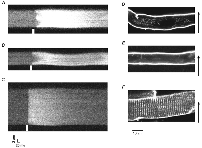
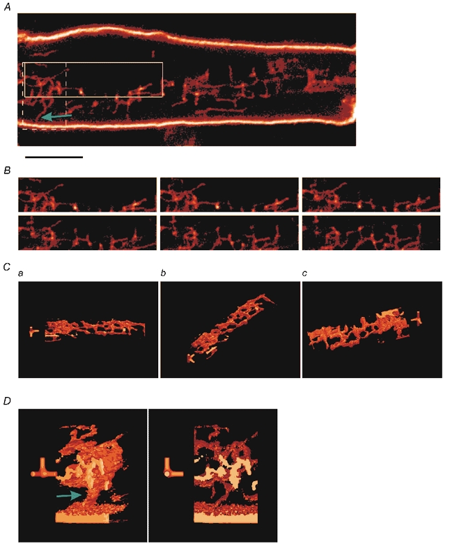
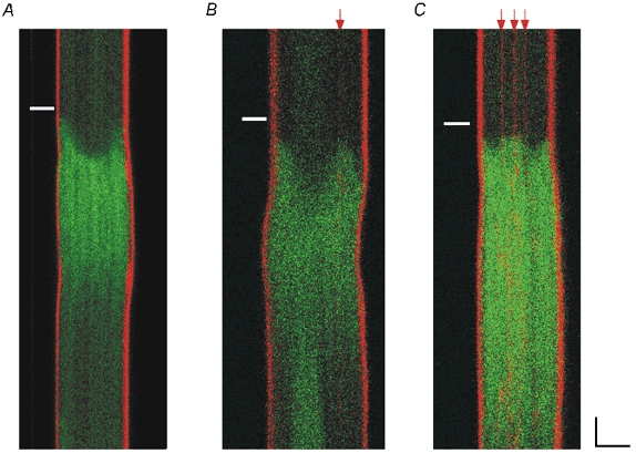
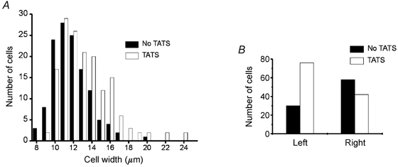
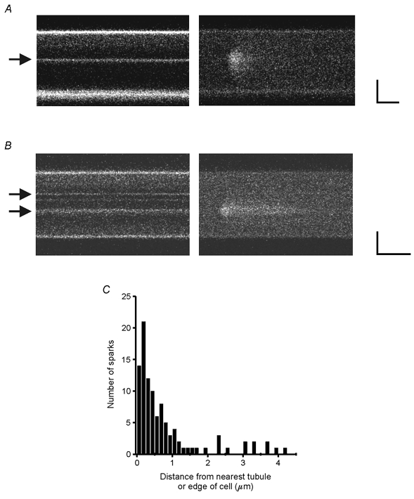
Cardiac atrial cells lack a regular system of transverse tubules like that in cardiac ventricular cells. Nevertheless, many atrial cells do possess an irregular internal transverse-axial tubular system (TATS). To investigate the possible role of the TATS in excitation-contraction coupling in atrial myocytes, we visualized the TATS (labelled with the fluorescent indicator, Di-8-ANEPPS) simultaneously with Ca2+ transients and/or Ca2+ sparks (fluo-4). In confocal transverse linescan images of field-stimulated cells, whole-cell Ca2+ transients had two morphologies: 'U-shaped' transients and irregular or 'W-shaped' transients with a varying number of points of origin of the Ca2+ transient. About half (54 %, n =289 cells, 13 animals) of the cells had a TATS. Cells with TATS had a larger mean diameter (13.2 +/- 2.8 microm) than cells without TATS (11.7 +/- 2.0 microm) and were more common in the left atrium (n = 206 cells; left atrium: 76 with TATS, 30 without TATS; right atrium: 42 with TATS, 58 without TATS). Simultaneous measurement of Ca2+ sparks and sarcolemmal structures showed that cells without TATS had U-shaped transients that started at the cell periphery, and cells with TATS had W-shaped transients that began simultaneously at the cell periphery and the TATS. Most (82 out of 102 from 31 cells) 'spontaneous' (non-depolarized) Ca2+ sparks occurred within 1 microm of a sarcolemmal structure (cell periphery or TATS), and 33 % occurred within 1 pixel (0.125 microm). We conclude that the presence of a sarcolemmal membrane either at the cell periphery or in the TATS in close apposition to the sarcoplasmic reticulum is required for the initiation of an evoked Ca2+ transient and for spontaneous Ca2+ sparks.


Figures

Figure 2
Figure 2. Relationship between TATS and the shape of the Ca2+ transient
Linescan images of Ca2+ transients in atrial (A and B) and ventricular (C) cells were taken along a direction transverse to the longitudinal axis of the cells (arrows). Ca2+ transients in atrial cells fell into two categories: those with numerous initiation points termed W-shaped (A) and U-shaped transients (B). Ventricular cells (C) exhibited nearly simultaneous activation across the cell. White bars denote the time of the field stimulation pulse. D-F show confocal images of Di-8-ANEPPS labelling of sarcolemmal membranes of atrial cells (D and E) and a ventricular cell (F). D shows a cell with a TATS with a prominent longitudinal component while the cell in E is almost devoid of TATS. The ventricular cell (F) shows a regular pattern of Di-8-ANEPPS labelling of T-tubules that extend from the surface and project radially into the cell.
Figure 1
Figure 1. Three-dimensional structure of TATS
A shows an optical section of a Di-8-ANEPPS-labelled atrial cell. The surface sarcolemma is heavily labelled as are finer internal membrane structures that are consistent with the previously described transverse-axial tubular system (TATS). The focal plane lies 7.25 μm from the bottom of the cell. Scale bar is 5 μm. B shows the region enclosed by the continuous box in A at different focal planes. The focal plane is 7.25 μm from the bottom of the cell (top left) and increases by 0.25 μm increments (left to right, top to bottom). C shows a 3-D reconstruction of the 5.5 μm thick section of the region in B. D shows a reconstruction of the 4.25 μm thick region in the dashed box in A. The green arrows in A and D show the connection between the surface sarcolemma and the TATS. The full-axis is along the axial direction and the half-axes are along longitudinal and transverse axes of the cell. In both C and D,the short axes are 1 μm long; the full-axis is 5.5 μm long in C and 4.25 μm in D.
Figure 3
Figure 3. The presence and location of sarcolemmal tubules determines the shape of the whole-cell Ca2+ transient
Cells were double-labelled with fluo-4 (green signal) and Di-8-ANEPPS (red signal) and scanned transversely. Red arrows highlight the positions of the sarcolemmal tubules. A, cell without TATS showing U-shaped transient. All cells without TATS had U-shaped transients (18 cells). B and C, cells with TATS usually (14 of 16 cells) had irregular, or W-shaped, transients and the points of the W corresponded to the locations of the tubules as marked by the Di-8-ANEPPS signal. White bars denote the time of the field stimulation pulse. Scale bars are 5 μm and 50 ms.
Figure 4
Figure 4. Cells with TATS are larger and the left atrium has more cells with TATS
A, histogram of cell widths with (□) and without (▪) TATS (289 cells from 13 animals). Cells with TATS had a slightly larger mean width than cells without TATS (13.2 vs. 11.7 μm, P < 0.0001, Mann-Whitney U test). B, a large majority of left atrial cells had a TATS (76 of 106 cells (72 %) had TATS, 30 (28 %) did not have TATS) but there was a slight preponderance of cells without TATS in the right atrium (42 of 100 cells (42 %) had TATS, 58 (58 %) did not have TATS). There is a statistically greater proportion of TATS in left atrial cells than in right atrial cells (P < 0.0001 by the Fisher exact test).
Figure 5
Figure 5. Spontaneous Ca2+ sparks occur on TATS
A and B show examples of spontaneous Ca2+ sparks in rat atrial cells double-labelled with Di-8-ANEPPS (left) and fluo-4 (right). Arrows highlight the location of the sarcolemmal tubules. Scale bars are 5 μm and 50 ms. C, histogram of distances between the position of the peak of the Ca2+ spark and the nearest membrane structure. Thirty-three per cent of the Ca2+ sparks were within 0.125 μm (1 pixel) of the nearest sarcolemmal structure and 80 % were within 1 μm.
Figure 6
Figure 6. Time-to-target plots distinguish between non-propagating and propagating Ca2+ release
A, transverse linescan image of a rat atrial cell. B, corresponding plot of the time to reach a target fluorescence level as a function of distance from the cell's lower edge (time-to-target plot). The concave-up time-to-target plot is consistent with non-propagating Ca2+ release from the edge of the cell and the passive diffusion of Ca2+ to the centre of the cell. C shows a linescan image from another cell and D its corresponding time-to-target plot. The linear time-to-target plot suggests that Ca2+ spreads to the centre of the cell as a propagated Ca2+ wave. The target fluorescence value is usually chosen to be at the midpoint of the baseline and peak fluorescence values. In cases where the fluorescence decreases rapidly from the surface (e.g. A), a lower target fluorescence value (∼20 % of peak fluorescence) is used to obtain more data points allowing a better determination of the shape of the time-to-target plot. Using both simulated and experimental linescans, we found that the target value does not change either the shape of the time-to-target plot or the calculated Ca2+ wave velocity. Colour scale bar ranges from 0 to 255 fluorescence units from black to yellow.

Similar articles

Cited by

References

    1. Berlin J. Spatiotemporal changes of Ca2+ during electrically evoked contractions in atrial and ventricular cells. Am J Physiol. 1995;269:H1165–1170. - PubMed
    1. Blatter LA, Hüser J, Rios E. Sarcoplasmic reticulum Ca2+ release flux underlying Ca2+ sparks in cardiac muscle. Proc Natl Acad Sci U S A. 1997;94:4176–4181. - PMC - PubMed
    1. Bridge JH, Ershler PR, Cannell MB. Properties of Ca2+ sparks evoked by action potentials in mouse ventricular myocytes. J Physiol. 1999;518:469–478. - PMC - PubMed
    1. Cannell MB, Cheng H, Lederer WJ. The control of calcium release in heart muscle. Science. 1995;268:1045–1049. - PubMed
    1. Cannell MB, Soeller C. Mechanisms underlying calcium sparks in cardiac muscle. J Gen Physiol. 1999;113:373–376. - PMC - PubMed

Publication types

Substances

LinkOut - more resources