. 2003 Mar 15;547(Pt 3):951-61.
doi: 10.1113/jphysiol.2002.038117. Epub 2003 Jan 24.

Troponin I in the murine myocardium: influence on length-dependent activation and interfilament spacing

Affiliations

Troponin I in the murine myocardium: influence on length-dependent activation and interfilament spacing

John P Konhilas et al. J Physiol. .

Abstract

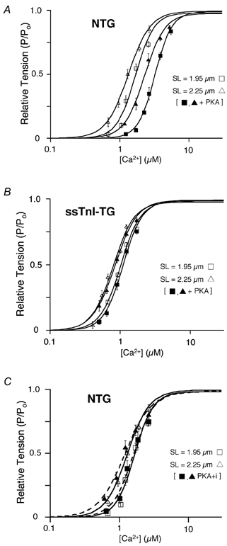
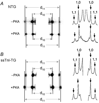
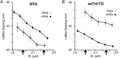
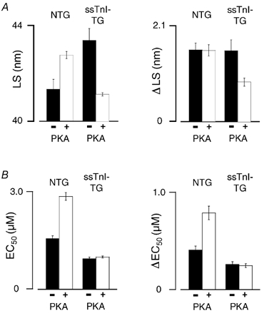
Cyclic AMP-dependent protein kinase (PKA) targets contractile proteins, troponin-I (TnI) and myosin binding protein C (MyBP-C) in the heart and induces a decrease in myofilament Ca2+ sensitivity. Yet, the effect of sarcomere length (SL) change on Ca2+ sensitivity (length-dependent activation: LDA) following PKA-dependent phosphorylation is not clear. To clarify the role of PKA-dependent phosphorylation of TnI and MyBP-C on LDA in the heart, we examined LDA in skinned myocytes from a non-transgenic (NTG) and a transgenic murine model in which the native cardiac isoform (cTnI) was completely replaced by the slow skeletal isoform of TnI (ssTnI-TG) lacking the phosphorylation sites for PKA, while retaining PKA sites on MyBP-C. In NTG myocytes, PKA treatment decreased Ca2+ sensitivity at each SL, but enhanced the impact of SL change on Ca2+ sensitivity. Despite a greater sensitivity to Ca2+ and a reduction in LDA, neither Ca2+ responsiveness nor LDA was affected by PKA treatment in ssTnI-TG myocytes. To determine whether the above observations could be explained by the lateral separation between thick and thin filaments, as suggested by others, we measured interfilament spacing by X-ray diffraction as a function of SL in skinned cardiac trabeculae in the passive state from both NTG and ssTnI-TG models before and following treatment with PKA. Phosphorylation by PKA increased lattice spacing at every SL in NTG trabeculae. However, the relationship between SL and myofilament lattice spacing in ssTnI-TG was markedly shifted downward to an overall decreased myofilament lattice spacing following PKA treatment. We conclude: (1) PKA-dependent phosphorylation enhances length-dependent activation in NTG hearts; (2) replacement of native TnI with ssTnI increases Ca2+ sensitivity of tension but reduces length-dependent activation; (3) MyBP-C phosphorylation by PKA does not alter calcium responsiveness and induces a decrease in myofilament lattice spacing at all sarcomere lengths and (4) length-dependent activation in the heart cannot be entirely explained by alterations in myofilament lattice spacing.


Figures

Figure 1
Figure 1. Ca2+-dependent tension development in cardiac myocytes from NTG and ssTnI-TG hearts
A and B, Ca2+-force relation of NTG (A) and ssTnI-TG (B) cardiac myocytes before (open symbols) and following (closed symbols) treatment with the catalytic subunit of PKA (3 μg protein (ml standard relaxing solution)−1) at SL = 2.25 μm (δ) and SL = 1.95 μm (□). C, Ca2+-force relation of NTG cardiac myocytes at two sarcomere lengths before (open symbols) and following (closed symbols) treatment with a PK inhibitor (PKI) added to PKA solution. Lines indicate the Hill fit to the data obtained at the indicated sarcomere length before and following PKA treatment.
Figure 2
Figure 2. Typical X-ray diffraction patterns obtained in skinned cardiac trabeculae from NTG and ssTnI-TG hearts before and following PKA treatment at SL = 2.10 μm
A, CCD images of the diffraction patterns. X-ray diffraction by the myofilament lattice gives rise to two sets of symmetrical, equatorial reflections, delineated by the continuous vertical lines. B, intensity profiles of the 2-dimensional CCD images shown in A. Arrows indicate the peak intensities, corresponding to the 1,0 and 1,1 reflections. These clearly resolved reflections or intensity peaks allowed lattice spacing to be determined within 0.25 % accuracy, equivalent to a myofilament lattice spacing resolution of 0.1 nm.
Figure 3
Figure 3. Average myofilament lattice spacing as a function of sarcomere length in skinned cardiac trabeculae from NTG and ssTnI-TG hearts (n = 8–10 for each group)
The relationships were obtained by averaging the data into 0.05 μm wide sarcomere length bins. A, relationship between lattice spacing and sarcomere length in NTG trabeculae prior to (○) and following (•) treatment with PKA. B, relationship between lattice spacing and sarcomere length in ssTnITG trabeculae prior to (○) and following (•) treatment with PKA. Each arrow indicates the sarcomere length at which the Ca2+-force relationships were determined.
Figure 4
Figure 4. Correlation between length change, Ca2+ sensitivity and lattice spacing with and without PKA treatment
A, comparison of lattice spacing (LS) and the effect of length change on lattice spacing (ΔLS in nm). Left, LS at SL = 2.1 mm in NTG and ssTnI-TG trabeculae treated with or without PKA as indicated. Right, the effect of length change on LS (ΔLS) in NTG and ssTnI-TG trabeculae treated with or without PKA. Error bars are ±s.e.m.B, comparison of Ca2+ sensitivity (EC50 in μmol l−1) and the effect of length change on Ca2+ sensitivity (ΔEC50 in μmol l−1). Left, ΔEC50 at SL = 2.1 mm in NTG and ssTnI-TG trabeculae treated with or without PKA as indicated. Right, the effect of length change on Ca2+ sensitivity (ΔEC50) in NTG and ssTnI-TG trabeculae treated with or without PKA.

Similar articles

Cited by

References

    1. Arteaga GM, Palmiter KA, Leiden JM, Solaro RJ. Attenuation of length dependence of calcium activation in myofilaments of transgenic mouse hearts expressing slow skeletal troponin I. J Physiol. 2000;526:541–549. - PMC - PubMed
    1. Backx PH, Gao WD, Azan-Backx MD, Marban E. Mechanism of force inhibition by 2,3-butanedione monoxime in rat cardiac muscle: roles of [Ca2+]i and cross-bridge kinetics. J Physiol. 1994;476:487–500. - PMC - PubMed
    1. Bagni MA, Cecchi G, Griffiths PJ, Maeda Y, Rapp G, Ashley CC. Lattice spacing changes accompanying isometric tension development in intact single muscle fibers. Biophys J. 1994;67:1965–1975. - PMC - PubMed
    1. Bremel RD, Weber A. Cooperation with actin filament in vertebrate skeletal muscle. Nat New Biol. 1972;238:97–101. - PubMed
    1. Brenner B, Yu LC. Equatorial x-ray diffraction from single skinned rabbit psoas fibers at various degrees of activation. Changes in intensities and lattice spacing. Biophys J. 1985;48:829–834. - PMC - PubMed

Publication types

LinkOut - more resources