

Response of human jaw muscles to axial stimulation of the incisor
- PMID: 12562936
- PMCID: PMC2342631
- DOI: 10.1113/jphysiol.2002.029579
Response of human jaw muscles to axial stimulation of the incisor
Abstract
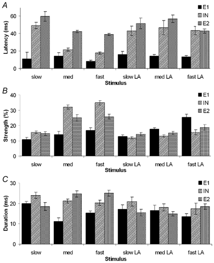
The role of periodontal mechanoreceptors (PMRs) in the reflex control of the jaw muscles has thus far been mainly derived from animal studies. To date, the work that has been done on humans has been limited and confined to orthogonal stimulation of the labial surface of the tooth. The purpose of this study was to investigate the response of the masseter and digastric muscles in humans to controlled axial stimulation of the upper left central incisor, both before and during a local anaesthetic block of the PMRs. Ten neurologically normal young adult females were tested, each on two separate occasions to confirm the reproducibility of the results. It was found that the reflex response in the masseter was modulated by the rate of rise of the stimulus used and, to a lesser degree, the level of background muscle activity. There was little detectable change in the activity of the digastric muscle under the tested conditions and what was found could be attributed to cross-talk with the masseter. The reflex responses obtained were significantly different between subjects; however retesting the same subject on a different occasion yielded similar results. The results indicate that the most common response of the masseter muscle to brisk axial stimulation of the incisor is a reflex inhibition at 20 ms, followed by a late excitation at 44 ms. However, it is possible that this late excitation could be due to delayed action potentials and hence be artefactual. As the application of a local anaesthetic block removed or significantly reduced both of these responses, it was concluded that they originated from the PMRs. Unlike during orthogonal stimulation, slowly rising stimuli did not produce any excitatory reflex activity. This indicated a difference in jaw reflexes to forces applied in different directions, possibly due to the activation of different receptor types when stimulating the tooth in either the orthogonal or axial directions.
Figures


Similar articles
-
Jaw movement alters the reaction of human jaw muscles to incisor stimulation.Exp Brain Res. 2005 Jul;164(2):165-76. doi: 10.1007/s00221-005-2239-4. Epub 2005 May 3. Exp Brain Res. 2005. PMID: 15868176
-
Response of human jaw muscles to axial stimulation of a molar tooth.Exp Brain Res. 2004 Nov;159(2):214-24. doi: 10.1007/s00221-004-1949-3. Epub 2004 Jul 6. Exp Brain Res. 2004. PMID: 15241573
-
Jaw reflexes evoked by mechanical stimulation of teeth in humans.J Neurophysiol. 1999 May;81(5):2156-63. doi: 10.1152/jn.1999.81.5.2156. J Neurophysiol. 1999. PMID: 10322056
-
[Masseter muscle reflex].Suom Hammaslaakarilehti. 1990 Aug 15;37(14):804-10. Suom Hammaslaakarilehti. 1990. PMID: 2244110 Review. Finnish. No abstract available.
-
Masseter reflexes modulated by pain.Mov Disord. 2002;17 Suppl 2:S45-8. doi: 10.1002/mds.10058. Mov Disord. 2002. PMID: 11836754 Review. No abstract available.
Cited by
-
The reciprocal jaw-muscle reflexes elicited by anterior- and back-tooth-contacts-a perspective to explain the control of the masticatory muscles.BDJ Open. 2020 Dec 17;6(1):27. doi: 10.1038/s41405-020-00056-z. BDJ Open. 2020. PMID: 33335091 Free PMC article.
-
Exteroceptive reflexes in jaw-closing muscle EMG during rhythmic jaw closing and clenching in man.Exp Brain Res. 2005 Apr;162(2):230-8. doi: 10.1007/s00221-004-2167-8. Epub 2004 Dec 10. Exp Brain Res. 2005. PMID: 15791466
-
Jaw movement alters the reaction of human jaw muscles to incisor stimulation.Exp Brain Res. 2005 Jul;164(2):165-76. doi: 10.1007/s00221-005-2239-4. Epub 2005 May 3. Exp Brain Res. 2005. PMID: 15868176
-
Response of human jaw muscles to axial stimulation of a molar tooth.Exp Brain Res. 2004 Nov;159(2):214-24. doi: 10.1007/s00221-004-1949-3. Epub 2004 Jul 6. Exp Brain Res. 2004. PMID: 15241573
-
Periodontal anaesthesia reduces common 8 Hz input to masseters during isometric biting.Exp Brain Res. 2006 Mar;169(3):326-37. doi: 10.1007/s00221-005-0144-5. Epub 2005 Nov 18. Exp Brain Res. 2006. PMID: 16328310
References
-
- Abbink JH, Van Der Bilt A, Bosman F, van der Glas HW, Erkelens CJ, Klaassen MF. Comparison of external load compensation during rhythmic arm movements and rhythmic jaw movements in humans. J Neurophysiol. 1999;82:1209–1217. - PubMed
-
- Anderson DJ. Measurement of stress in mastication II. J Dent Res. 1956;35:671–673. - PubMed
-
- Brinkworth RSA, Savundra AW, Türker KS, Yeo PLM, Linke IR, Sampson W. Response of human jaw muscles to axial stimulation. Proc Aust Neurosci Soc. 2002;13:121.
-
- Brodin P, Miles TS, Türker KS. Simple reaction-time responses to mechanical and electrical stimuli in human masseter muscle. Arch Oral Biol. 1993a;38:221–226. - PubMed
Publication types
MeSH terms
LinkOut - more resources
Full Text Sources
Research Materials