

Glucocorticoid exposure at the dose used clinically alters cytoskeletal proteins and presynaptic terminals in the fetal baboon brain
- PMID: 12562943
- PMCID: PMC2342613
- DOI: 10.1113/jphysiol.2002.025700
Glucocorticoid exposure at the dose used clinically alters cytoskeletal proteins and presynaptic terminals in the fetal baboon brain
Abstract
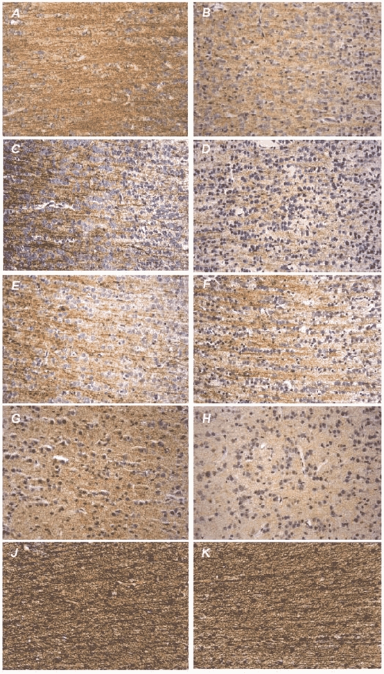
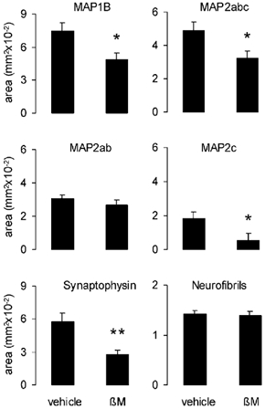
Glucocorticoids have been used for 30 years to accelerate fetal lung maturation in human pregnancy at risk of preterm delivery. Exposure to inappropriate levels of steroid, however, leads to altered maturation of the cardiovascular, metabolic and central nervous systems. The effects of betamethasone on neuronal development and function were determined in the fetal baboon brain by examination of cytoskeletal microtubule associated proteins (MAPs) and the presynaptic marker protein synaptophysin. At 0.73 gestation, commencing 28 weeks of gestation, pregnant baboons received four doses of saline (n = 8) or 87.5 microg (kg body weight)(-1) betamethasone I.M. (n = 7) 12 h apart. This dose is equivalent to 12 mg betamethasone administered daily over two consecutive days to a 70 kg woman. Baboons underwent Caesarean section 12 h after the last injection. Paraffin sections of the fetal neocortex and the underlying white matter were labelled immunohistochemically against MAP1B, MAP2abc, MAP2ab and synaptophysin and stained histochemically with hematoxylin-eosin and silver. Tissue staining was quantified morphometrically. Betamethasone exposure resulted in decreased immunoreactivity (IR) of MAP1B by 34.3 % and MAP2abc by 34.1 % (P < 0.05). Loss of MAP2 IR was due to loss of IR of the juvenile isoform MAP2c (P < 0.05). MAP1B and MAP2c are involved in neuritogenesis and neuronal plasticity. Synaptophysin IR was reduced by 51.8 % (P < 0.01). These changes might reflect functional neuronal disturbances because they were not accompanied by an alteration of the density of neurofibrils or neuronal necrosis. These results are in agreement with earlier findings of alterations of cytoskeletal proteins and presynaptic terminals in the fetal sheep brain after betamethasone infusion directly to the fetus and support a common effect of inappropriate fetal exposure to glucocorticoids on neuronal cytoskeleton and synapses in mammalian species.
Figures
References
-
- Antonow-Schlorke I, Kühn B, Müller T, Schubert H, Sliwka U, Nathanielsz PW, Schwab M. Effect of antenatal betamethasone treatment on density of presynaptic terminals in the fetal sheep. Neurosci Lett. 2001;297:147–150. - PubMed
-
- Arnold SE, Trojanowski JQ. Human fetal hippocampal development: II The neuronal cytoskeleton. J Comp Neurol. 1996;367:293–307. - PubMed
-
- Barker DJP. Mothers, Babies and Health in Later Life. Edinburgh: Churchill Livingstone; 1998.
-
- Bonfoco E, Ceccatelli S, Manzo L, Nicotera P. Colchicine induces apoptosis in cerebellar granule cells. Exp Cell Res. 1995;218:189–200. - PubMed
Publication types
MeSH terms
Substances
Grants and funding
LinkOut - more resources
Full Text Sources
Other Literature Sources
Medical