

Augmentation of coronary conductance in adult sheep made anaemic during fetal life
- PMID: 12562949
- PMCID: PMC2342629
- DOI: 10.1113/jphysiol.2002.023283
Augmentation of coronary conductance in adult sheep made anaemic during fetal life
Abstract
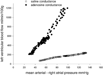
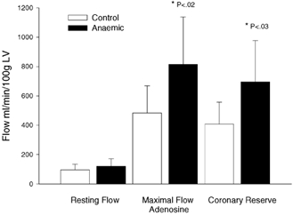
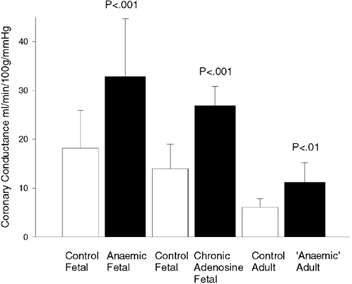
Maximal coronary conductance with adenosine in anaemic fetal sheep is twice that of non-anaemic fetuses. To investigate whether this increase in conductance persists into adulthood we studied twin sheep as fetuses and again as adults. Nine anaemic fetuses (118 days gestation) underwent isovolaemic haemorrhage for 18.0 +/- 4.6 days (means +/- S.D.) during which time the haematocrit was reduced from 39.9 +/- 5.2 % to 16.3 +/- 3.4 % and oxygen content from 8.6 +/- 1.3 to 2.3 +/- 0.2 ml dl-1. At 138 days the anaemic fetuses were transfused; at delivery the haematocrit was 29.3 +/- 6.8 % compared to nine control fetuses in which the haematocrit was 38.5 +/- 4.3 %. The weight at delivery was 3.5 +/- 0.36 kg in the anaemic fetuses vs. 4.2 +/- 0.83 kg in controls. Twenty-eight weeks later, we placed an occluder on the descending thoracic aorta and inferior vena cava, a flow probe around the proximal left circumflex coronary artery, and catheters in the left atrial appendage, jugular and carotid vessels. Maximal coronary conductance was determined in the adults by recording coronary blood flow as driving pressure was altered by inflating the occluders while adenosine was infused into the left atrium. Right atrial, left atrial, systolic and mean arterial pressures, systemic vascular resistance and haematocrit were not different between 'in utero anaemic' and control adults. The adults that were anaemic in utero weighed less than the controls 39.4 +/- 4.6 kg vs. 45.0 +/- 5.6 kg. Maximal conductance was greater in the adults that were anaemic in utero: 11.2 +/- 4.0 ml min(-1) (100 g)(-1) mmHg-1 as compared to 6.1 +/- 1.8 ml min(-1) (100 g)(-1) mmHg(-1) in the controls. Vascular reactivity of the mesenteric arteries was not different. These data suggest that coronary conductance can be modified in utero by anaemia (high flow and hypoxaemia) and that the remodelled coronary tree persists to adulthood.
Figures
References
-
- Bache R, Alyono D, Sublett E, Dai X. Myocardial blood flow in left ventricular hypertrophy developing in young and adult dogs. American Journal of Physiology. 1986;251:H949–956. - PubMed
-
- Barker DJP, editor. Fetal Origins of Cardiovascular and Lung Disease. New York: Marcel Dekker, Inc.; 2000. pp. V–VII. Preface.
-
- Bishop S, Powell P, Hasebe N, Shen Y, Patrick T, Hittinger L, Vatner S. Coronary vascular morphology in pressure-overload left ventricular hypertrophy. Journal of Molecular and Cellular Cardiology. 1996;28:141. - PubMed
-
- Dalshaug G, Scholz T, Smith O, Bedell K, Caldarone C, Segar J. Effects of gestational age on myocardial blood flow and coronary flow reserve in pressure-loaded ovine fetal hearts. American Journal of Physiology - Heart and Circulatory Physiology. 2002;282:H1359–1369. - PubMed
-
- Davis L, Hohimer A. Hemodynamics and organ blood flow in fetal sheep subjected to chronic anemia. American Journal of Physiology. 1991;261:R1542–1548. - PubMed
Publication types
MeSH terms
Substances
Grants and funding
LinkOut - more resources
Full Text Sources
Medical
Miscellaneous