. 2003 Feb 15;547(Pt 1):133-45.
doi: 10.1113/jphysiol.2002.030676. Epub 2002 Dec 13.

Chronic hypoxia causes angiogenesis in addition to remodelling in the adult rat pulmonary circulation

Affiliations

Chronic hypoxia causes angiogenesis in addition to remodelling in the adult rat pulmonary circulation

Katherine Howell et al. J Physiol. .

Abstract

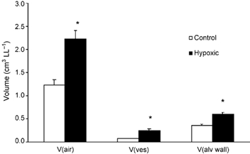
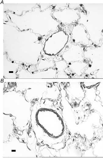
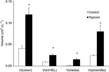
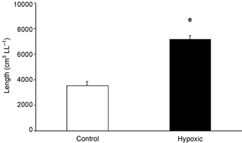
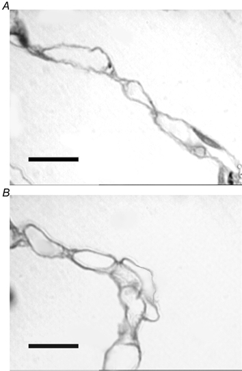
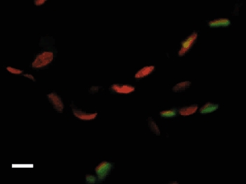
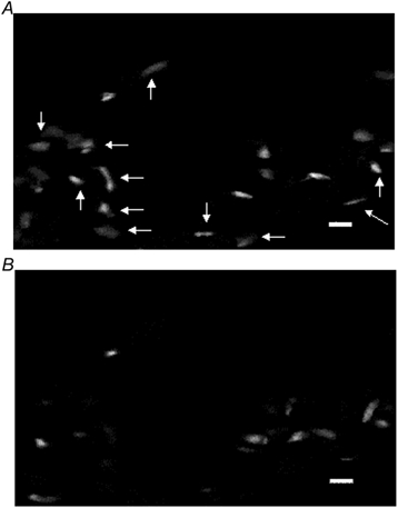
Chronic hypoxia caused by migration of native sea-level dwellers to high altitude or chronic lung disease leads to the development of increased pulmonary vascular resistance and pulmonary hypertension. This altitude-induced hypertension offers no obvious benefit and may indeed be maladaptive. A major mechanism thought to contribute to the development of pulmonary hypertension is hypoxia-induced loss of small blood vessels, sometimes termed rarefaction or pruning. More recent evidence caused us to question this widely accepted concept including the potent angiogenic effect of chronic hypoxia in all other vascular beds and the demonstration that new vessels can form in the pulmonary circulation when stimulated by chronic infection and lung resection. We tested the hypothesis that chronic environmental hypoxia causes angiogenesis in the adult pulmonary circulation by using stereological techniques combined with confocal microscopy to examine the resultant changes in pulmonary vascular structure in rats. We found that chronic hypoxia resulted in increased total pulmonary vessel length, volume, endothelial surface area and number of endothelial cells in vivo. This is the first reported demonstration of hypoxia-induced angiogenesis in the mature pulmonary circulation, a structural adaptation that may have important beneficial consequences for gas exchange. These findings imply that we must revise the widely accepted paradigm that hypoxia-induced loss of small vessels is a key structural change contributing to the development of pulmonary hypertension in high altitude adaptation and chronic lung disease.


Figures

Figure 1
Figure 1. Mean (± s.e.m.) volume of intra-acinar sub-compartments for control (n = 7) and hypoxic (n = 6) animals
V(air), volume of intra-acinar airspaces; V(ves), volume of intra-acinar pulmonary blood vessels excluding capillaries; V(alv wall), volume of intra-acinar alveolar wall including capillaries. *Significant difference from controls (P < 0.05, t test).
Figure 2
Figure 2. Photomicrographs of intra-acinar blood vessels in control and hypoxic lungs
Both images were taken from semi-thin resin sections stained with Toluidine Blue. A, photomicrograph of intra-acinar vessel from control lungs. Vessel is typically thin walled with no medial layer and a single elastic lamina. B, photomicrograph of a typically remodelled intra-acinar vessel from chronically hypoxic lungs. Vessel demonstrates medial thickening, with both an internal and an external elastic lamina present. Scale bars indicate 20 µm.
Figure 3
Figure 3. Mean (± s.e.m.) volume of intra-acinar vessel lumen, tunica intima plus internal elastic lamina together, tunica media and tunica adventitia in intra-acinar pulmonary blood vessels in the left lungs of control (n = 7) and hypoxic (n = 6) animals
V(lumen), volume of intra-acinar blood vessel lumen; V(int + IEL), volume of tunica intima plus internal elastic lamina considered together (see Methods for details); V(media), volume of tunica media; V(adventitia), volume of tunica adventitia. *Significant difference from control animals (P < 0.001, t test).
Figure 4
Figure 4. Mean (± s.e.m.) intra-acinar vessel length per left lung in control and hypoxic animals
*Significant difference from controls (P < 0.01, t test).
Figure 5
Figure 5. Images of alveolar walls taken from semi-thin (1–2 µm) sections stained with Toluidine Blue
A, alveolar wall taken from control lung with numerous capillaries discernible within the alveolar wall. B, alveolar wall taken from chronically hypoxic lung tissue. Some capillaries appear to protrude from alveolar wall into alveolar lumen, a pattern not seen in control lungs. Scale bars indicate 10 µm.
Figure 6
Figure 6. Photomicrograph showing a confocally acquired image of control lung tissue
Nuclei were stained with Propidium Iodide (red) while endothelial cells were stained using anti-VEGFR-2 antibody visualised using FITC (green). Scale bar represents 10 µm.
Figure 7
Figure 7. Photomicrographs showing confocally acquired images of endothelial cells stained immunofluorescently with anti-VEGFR-2 antibody labelled with FITC
The number of endothelial cells seen in the control lung section (A) is less than that observed in the hypoxic lung section (B). Scale bars represent 10 µm.

References

    1. Abraham AS, Kay JM, Cole RB, Pincock AC. Haemodynamic and pathological study of the effect of chronic hypoxia and subsequent recovery of the heart and pulmonary vasculature of the rat. Cardiovasc Res. 1971;5:95–102. - PubMed
    1. Ausprunk DH, Folkman J. Migration and proliferation of endothelial cells in preformed and newly formed blood vessels during tumor angiogenesis. Microvasc Res. 1977;14:53–65. - PubMed
    1. Bolender RP, Hyde DM, Dehoff RT. Lung morphometry: a new generation of tools and experiments for organ, tissue, cell, and molecular biology. Am J Physiol. 1993;265:L521–548. - PubMed
    1. Brogi E, Schatteman G, Wu T, Kim EA, Varticovski L, Keyt B, Isner JM. Hypoxia-induced paracrine regulation of vascular endothelial growth factor receptor expression. J Clin Invest. 1996;97:469–476. - PMC - PubMed
    1. Christou H, Yoshida A, Arthur V, Morita T, Kourembanas S. Increased vascular endothelial growth factor production in the lungs of rats with hypoxia-induced pulmonary hypertension. Am J Respir Cell Mol Biol. 1998;18:768–776. - PubMed

Publication types

MeSH terms

LinkOut - more resources