

A mechanism underlying the sexually dimorphic ACTH response to lipopolysaccharide in rats: sex steroid modulation of cytokine binding sites in the hypothalamus
- PMID: 12562959
- PMCID: PMC2342603
- DOI: 10.1113/jphysiol.2002.032169
A mechanism underlying the sexually dimorphic ACTH response to lipopolysaccharide in rats: sex steroid modulation of cytokine binding sites in the hypothalamus
Abstract
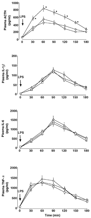
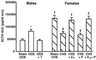
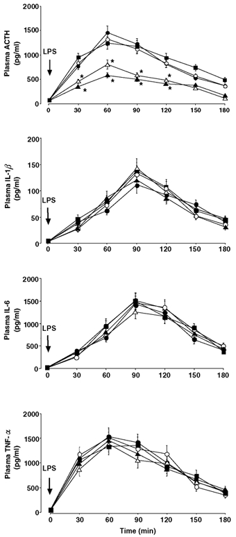
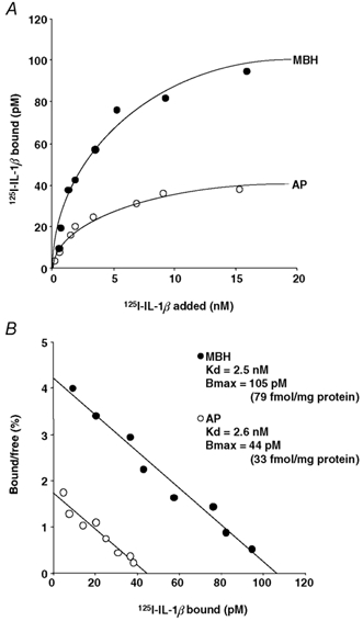
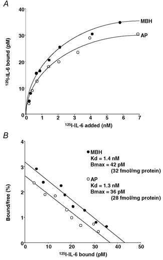
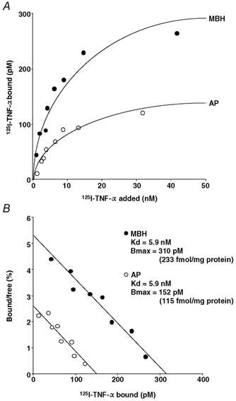
It is well established that the hypothalamic-pituitary-adrenal responses to immune stressors are sexually dimorphic in rodents (females > males), but the underlying mechanism is still unclear. To investigate the mechanism, in this study we examined whether the sex steroid environment affects the following variables in male and female rats: (1) plasma levels of ACTH, interleukin (IL)-1beta, IL-6 and tumour necrosis factor-alpha (TNF-alpha) after systemic lipopolysaccharide (LPS) administration; (2) static concentrations of corticotropin-releasing hormone (CRH) and arginine vasopressin (AVP) in the mediobasal hypothalamus (MBH) and those of ACTH in the anterior pituitary (AP); and (3) the binding characteristics of IL-1beta, IL-6 and TNF-alpha in the MBH and AP. LPS-induced ACTH release was significantly higher in female than in male rats, and this sexual difference was abolished by performing gonadectomy in both sexes. Administration of physiological doses of testosterone and oestradiol to gonadectomized males and females, respectively, restored the altered ACTH responses to normal. Changes in the sex steroid milieu did not affect plasma cytokine responses to LPS, tissue contents of CRH, AVP and ACTH, or the IL-6 binding characteristics in the MBH and AP. However, the number of IL-1beta and TNF-alpha binding sites, but not their binding affinities, in the MBH showed significant changes according to altered sex hormone milieu, in the same direction as the LPS-induced ACTH response. These results suggest that the hypothalamic sensitivity to peripheral IL-1beta and TNF-alpha may be an important mechanism underlying the sexually dimorphic ACTH response to LPS in rats.
Figures
References
-
- Ban E, Milon G, Prudhomme N, Fillion G, Haour F. Receptors for interleukin-1 (α and β) in mouse brain: mapping and neuronal localization in hippocampus. Neuroscience. 1991;43:21–30. - PubMed
-
- Bohler HC, Zoeller RT, King JC, Rubin BS, Weber R, Merriam GR. Corticotropin releasing hormone mRNA is elevated on the afternoon of proestrus in the parvocellular paraventricular nuclei of the female rat. Mol Brain Res. 1990;8:259–262. - PubMed
-
- Broad KD, Keverne EB, Kendrick KM. Corticotropin releasing factor mRNA expression in the sheep brain during pregnancy, parturition and lactation and following exogenous progesterone and oestrogen treatment. Mol Brain Res. 1995;29:310–316. - PubMed
-
- Broadwell RD, Brightman MW. Entry of peroxidase into neurons of the central and peripheral nervous systems from extracerebral and cerebral blood. J Comp Neurol. 1976;166:257–283. - PubMed
Publication types
MeSH terms
Substances
LinkOut - more resources
Full Text Sources
Miscellaneous