Review
. 2003 Feb 15;547(Pt 1):21-33.
doi: 10.1113/jphysiol.2002.031534. Epub 2003 Jan 10.

Endothelial nitric oxide synthase, caveolae and the development of atherosclerosis

Affiliations
Review

Endothelial nitric oxide synthase, caveolae and the development of atherosclerosis

Philip W Shaul. J Physiol. .

Abstract

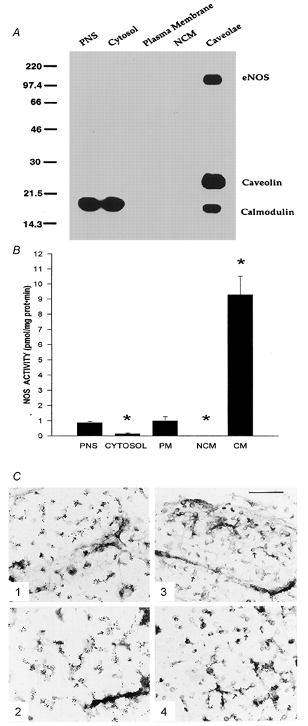
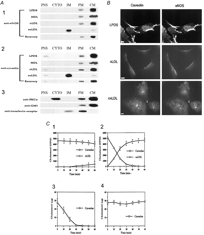
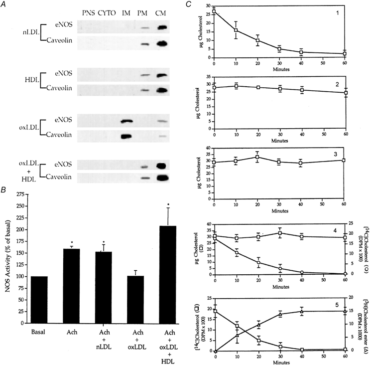
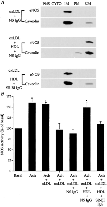
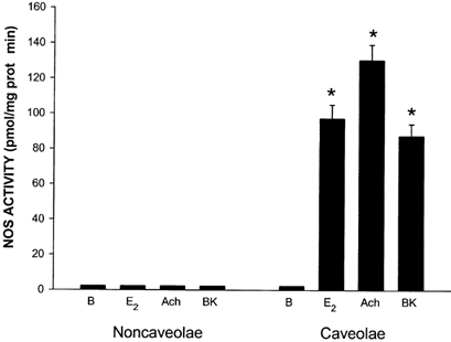
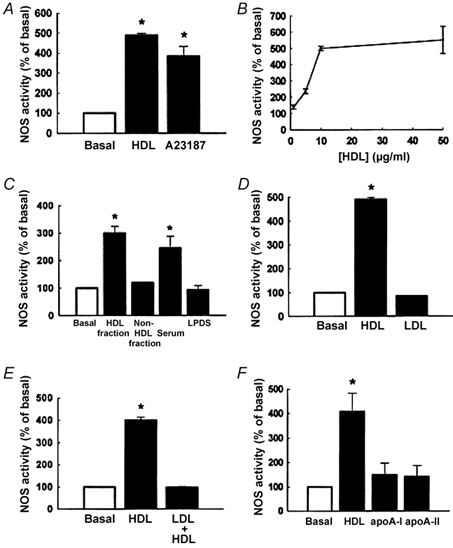
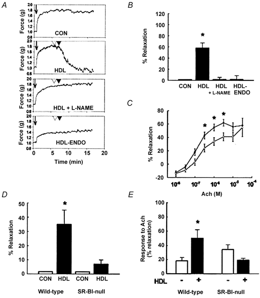
Early hypercholesterolaemia-induced vascular disease is characterized by an attenuated capacity for endothelial production of the antiatherogenic molecule nitric oxide (NO), which is generated by endothelial NO synthase (eNOS). In recent studies we have determined the impact of lipoproteins on eNOS subcellular localization and action, thereby providing a causal link between cholesterol status and initial abnormalities in endothelial function. We have demonstrated that eNOS is normally targeted to cholesterol-enriched caveolae where it resides in a signalling module. Oxidized low density lipoprotein (LDL; oxLDL) causes displacement of eNOS from caveolae by binding to endothelial cell CD36 receptors and by depleting caveolae cholesterol content, resulting in the disruption of eNOS activation. The adverse effects of oxLDL are fully prevented by high density lipoprotein (HDL) via binding to scavenger receptor BI (SR-BI), which is colocalized with eNOS in endothelial caveolae. This occurs through the maintenance of caveolae cholesterol content by cholesterol ester uptake from HDL. As importantly, HDL binding to SR-BI causes robust stimulation of eNOS activity in endothelial cells, and this process is further demonstrable in isolated endothelial cell caveolae. HDL also enhances endothelium- and NO-dependent relaxation in aortae from wild-type mice, but not in aortae from homozygous null SR-BI knockout mice. Thus, lipoproteins have potent effects on eNOS function in caveolae via actions on both membrane cholesterol homeostasis and the level of activation of the enzyme. These processes may be critically involved in the earliest phases of atherogenesis, which recent studies suggest may occur during fetal life.


Figures

Figure 1
Figure 1. eNOS is localized to endothelial cell caveolae
A, immunoblot analysis for eNOS, caveolin-1 and calmodulin in subcellular fractions from endothelial cells. Samples of postnuclear supernatant (PNS), cytosol, plasma membrane (PM), noncaveolae membrane (NCM) and caveolae membrane (CM) were evaluated. B, NOS enzymatic activity in subcellular fractions from endothelial cells. [3H]-l-arginine conversion to [3H]-l-citrulline was measured in the presence of excess substrate, cofactors, calcium and calmodulin. Enzymatic activity was undetectable in NCM. Values are means ± s.e.m., n = 4-6, * P < 0.05versus plasma membrane. C, localization of caveolin and eNOS in plasma membranes by immunoelectron microscopy. Immunogold labelling was performed using antibody to caveolin-1 in fibroblasts (panel 1) and endothelial cells (panel 2), and antibody to eNOS in fibroblasts (panel 3) and endothelial cells (panel 4). Caveolae (arrows) are evident in both cell types. Bar = 0.45 µm. Modified from Shaul et al. (1996).
Figure 2
Figure 2. Oxidized LDL displaces eNOS from endothelial cell caveolae due to depletion of caveolae cholesterol
A, oxidized LDL (oxLDL) but not lipoprotein-deficient serum (LPDS), HDL or nLDL exposure (60 min) alters the subcellular distribution of eNOS and caveolin. Additional studies were performed in oxLDL-treated cells that were washed and incubated for an additional 120 min in LPDS only (Recovery). Samples of postnuclear supernatant (PNS), cytosol (CYTO), intracellular membranes (IM), plasma membrane (PM) and caveolae membrane (CM) were isolated, and immunoblot analysis was performed for eNOS (panel 1) and caveolin-1 (panel 2). Control experiments in oxLDL-treated cells included immunoblot analyses for the caveolae resident proteins protein kinase C and GM1, and for transferrin receptors which are found in coated pits (panel 3). B, oxLDL but not LPDS or nLDL induces eNOS and caveolin to colocalize in an internal membrane compartment. Cells were incubated for 60 min, fixed and processed for double label immunofluorescence. C, oxLDL depletes caveolae of cholesterol. For panels 1 and 2, endothelial cells were labelled with [3H]acetate for 18 h at 37 °C, washed and incubated with nLDL (panel 1) or oxLDL (panel 2) for 0–60 min at 37 °C. The medium was collected, and the nLDL or oxLDL was isolated by centrifugation. The cells were washed, processed to isolate caveolae and [3H]cholesterol was extracted and measured. For panels 3 and 4, unlabelled cells were incubated with oxLDL (panel 3) or HDL (panel 4) for 0–60 min at 37 °C. The cells were washed, caveolae were isolated, and the amount of total cholesterol associated with caveolae was determined. Values are means ± s.e.m., n = 8. Modified from Blair et al. (1999).
Figure 3
Figure 3. HDL prevents oxLDL-induced inhibition of eNOS localization and activation in caveolae
A, endothelial cells were treated for 60 min with nLDL, HDL, or oxLDL, or with oxLDL followed by HDL for an additional 15 min. Samples of postnuclear supernatant (PNS), cytosol (CYTO), intracellular membranes (IM), plasma membrane (PM) and caveolae membrane (CM) were isolated, and immunoblot analysis was performed for eNOS and caveolin-1. B, after treating endothelial cells as described above, NOS activity was evaluated in intact cells by measuring [3H]l-arginine conversion to [3H]l-citrulline over 15 min in the absence of exogenous stimulation (Basal) or in the presence of acetylcholine (Ach, 10−6m). Values are means ± s.e.m., n = 4, * P < 0.05versus basal. C, HDL maintains the sterol content of caveolae. Endothelial cells were incubated with oxLDL (panel 1) or HDL (panel 2) for 0–60 min at 37 °C. For panel 3, cells were pretreated with oxLDL and then HDL was added for an additional 15 min. The cells were fractionated to isolate caveolae and the mass of cholesterol associated with caveolae was determined. In panel 4, cells were radiolabelled with [14C]acetate for 18 h, and HDL and oxLDL were added simultaneously for 0–60 min. The cells were processed to measure the amount of [14C]cholesterol associated with caveolae and the mass of cholesterol associated with caveolae. In panel 5, cellular cholesterol pools were radiolabelled as described for panel 4, and HDL was labelled with [3H]cholesterol ester. [3H]HDL and oxLDL were incubated with cells as described for panel 4, and the amount of [14C]cholesterol and [3H]cholesterol associated with caveolae was determined. Values are means ± s.e.m., n = 3. Modified from Uittenbogaard et al. (2000).
Figure 4
Figure 4. SR-BI mediates the effects of HDL on eNOS localization and activation in caveolae
A, endothelial cells were treated for 1 h with oxLDL, or oxLDL and HDL in the presence of nonspecific IgG or SR-BI IgG. Samples of postnuclear supernatant (PNS), cytosol (CYTO), intracellular membranes (IM), plasma membrane (PM) and caveolae membrane (CM) were isolated, and immunoblot analysis was performed for eNOS and caveolin-1. Representative data from four independent experiments are shown. B, after treating endothelial cells as described above, NOS activity was evaluated in intact cells by measuring [3H]-l-arginine conversion to [3H]-l-citrulline over 15 min in the absence of exogenous stimulation (Basal) or in the presence of acetylcholine (Ach, 10−6m). Values are means ± s.e.m., n = 4, * P < 0.05versus basal. Reprinted with permission (Uittenbogaard et al. 2000).
Figure 5
Figure 5. eNOS is localized to a functional signalling module in endothelial cell caveolae
[3H]-l-arginine conversion to [3H]-l-citrulline was measured in isolated noncaveolae and caveolae fractions of endothelial cell plasma membranes, in the absence (basal, B) or presence of 10−8m oestradiol (E2), 10−6m acetylcholine (Ach), or 10−6m bradykinin (BK). Values are means ± s.e.m., n = 4-6, * P < 0.05vs. basal. Modified from Chambliss et al. (2000).
Figure 6
Figure 6. HDL activates eNOS in endothelial cells
A, [3H]l-arginine conversion to [3H]l-citrulline was measured under basal conditions or in the presence of HDL or A23187. B, the dose-response to HDL was also assessed. C, eNOS activation was evaluated under basal conditions or in the presence of the HDL fraction or the non-HDL fraction of human serum, or whole serum, or lipoprotein-deficient serum (LPDS) added at equal volumes. D, the responses to HDL and LDL were compared. E, eNOS activation by HDL was assessed in the absence or presence of excess LDL. F, eNOS activation was determined in the presence of HDL or recombinant apoA-I or lipid-free apoA-II purified from human plasma. Values are means ± s.e.m., n = 3-6, * P < 0.05vs. basal. Reprinted with permission from Yuhanna et al. (2001).
Figure 7
Figure 7. HDL enhances endothelial NO production in aortae from wild-type mice, but not in aortae from homozygous SR-BI null mutant mice
A, following precontraction of rings of thoracic aortae from wild-type CD-1 mice with phenylephrine (arrow), direct relaxation responses to control buffer (CON), HDL at 10 µg ml−1 (open arrowhead) or 25 µg ml−1 (filled arrowhead), HDL with prior l-NAME treatment, or HDL with endothelium-denuded (- ENDO) rings were evaluated. B, cumulative findings (means ± s.e.m., for maximal relaxation to 25 µg ml−1 HDL in n = 10, 10, 3 and 5 studies, respectively, which were performed as in A. C, HDL augments the relaxation response to acetylcholine (Ach) in aortae from wild-type CD-1 mice. Studies were performed in control rings (open circles) and rings exposed to HDL (filled circles). D, the direct effects of control buffer (CON) or HDL were tested on phenylephrine-contracted rings of thoracic aortae from wild-type 129/C57BL/6 mice (SR-BI+/+) or homozygous null mutant mice (SR-BI-/-) mice. E, rings of thoracic aortae from SR-BI+/+ or SR-BI-/- mice were precontracted with phenylephrine, and the response to Ach was determined before and after exposure to HDL. In C, D and E, values are means ± s.e.m., and a minimum of 4 rings from 4 different mice were studied in each group. * P < 0.05vs. no HDL. Reprinted with permission from Yuhanna et al. (2001).

Similar articles

Cited by

References

    1. Anderson RG, Kamen BA, Rothberg KG, Lacey SW. Potocytosis: sequestration and transport of small molecules by caveolae. Science. 1992;255:410–411. - PubMed
    1. Blair A, Shaul PW, Yuhanna IS, Conrad PA, Smart EJ. Oxidized low density lipoprotein displaces endothelial nitric-oxide synthase (eNOS) from plasmalemmal caveolae and impairs eNOS activation. J Biol Chem. 1999;274:32512–32519. - PubMed
    1. Cayatte AJ, Palacino JJ, Horten K, Cohen RA. Chronic inhibition of nitric oxide production accelerates neointima formation and impairs endothelial function in hypercholesterolemic rabbits. Arterioscler Thromb. 1994;14:753–759. - PubMed
    1. Chambliss KL, Yuhanna IS, Mineo C, Liu P, German Z, Sherman TS, Mendelsohn ME, Anderson RG, Shaul P. Estrogen receptor alpha and endothelial nitric oxide synthase are organized into a functional signaling module in caveolae. Circ Res. 2000;87:E44–52. - PubMed
    1. Chang WJ, Rothberg KG, Kamen BA, Anderson RG. Lowering the cholesterol content of MA104 cells inhibits receptor-mediated transport of folate. J Cell Biol. 1992;118:63–69. - PMC - PubMed

Publication types

LinkOut - more resources