. 2003 Feb 15;547(Pt 1):125-32.
doi: 10.1113/jphysiol.2002.031450. Epub 2002 Dec 20.

Interactions between neutrophils and macrophages promote macrophage killing of rat muscle cells in vitro

Collaborators, Affiliations

Interactions between neutrophils and macrophages promote macrophage killing of rat muscle cells in vitro

Hal X Nguyen et al. J Physiol. .

Abstract

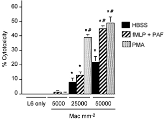
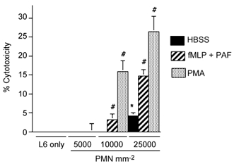
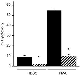
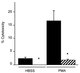
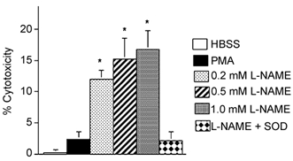
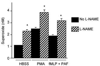
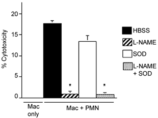
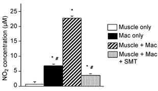
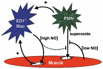
Current evidence indicates that the physiological functions of inflammatory cells are highly sensitive to their microenvironment, which is partially determined by the inflammatory cells and their potential targets. In the present investigation, interactions between neutrophils, macrophages and muscle cells that may influence muscle cell death are examined. Findings show that in the absence of macrophages, neutrophils kill muscle cells in vitro by superoxide-dependent mechanisms, and that low concentrations of nitric oxide (NO) protect against neutrophil-mediated killing. In the absence of neutrophils, macrophages kill muscle cells through a NO-dependent mechanism, and the presence of target muscle cells causes a three-fold increase in NO production by macrophages, with no change in the concentration of inducible nitric oxide synthase. Muscle cells that are co-cultured with both neutrophils and macrophages in proportions that are observed in injured muscle show cytotoxicity through a NO-dependent, superoxide-independent mechanism. Furthermore, the concentration of myeloid cells that is necessary for muscle killing is greatly reduced in assays that use mixed myeloid cell populations, rather than uniform populations of neutrophils or macrophages. These findings collectively show that the magnitude and mechanism of muscle cell killing by myeloid cells are modified by interactions between muscle cells and neutrophils, between muscle cells and macrophages and between macrophages and neutrophils.


Figures

Figure 1
Figure 1. Macrophages lyse muscle cells in vitro
Results of chromium release cytotoxicity assays of rat ED1+ macrophage killing of L6 myotubes. Bars = s.e.m.n = 6 for each group. * Significantly different from L6 muscle cells only under same culture conditions at P < 0.01. # Significantly different from macrophage and muscle co-cultures with HBSS in the absence of exogenous activators. Mac = macrophages.
Figure 2
Figure 2. Neutrophils lyse muscle cells in vitro
Results of chromium release cytotoxicity assays of rat neutrophil killing of L6 myotubes. Bars = s.e.m.n = 6 for each group. * Significantly different from muscle cells only under same culture conditions at P < 0.01. # Significantly different from neutrophil and muscle co-cultures with HBSS in the absence of exogenous activators. PMN = neutrophils.
Figure 3
Figure 3. NOS inhibition prevents muscle lysis in macrophage co-cultures
Cultures contained 25 000 macrophages mm−2. Cultures received no exogenous activator (HBSS) or were activated with PMA. Black bar = no l-NAME. Hatched bar = 150 µm l-NAME. * Significantly different from no l-NAME at P < 0.01. Bars = s.e.m.n = 6 for each group.
Figure 4
Figure 4. Superoxide dismutase decreases muscle lysis in neutrophil co-cultures
Cultures contained 10 000 neutrophils mm−2. Cultures received no exogenous activator (HBSS) or were activated with PMA. Black bar = no SOD. Hatched bar = 500 U ml−1 SOD. * Significantly different from no SOD at P < 0.01. Bars = s.e.m.n = 6 for each group.
Figure 5
Figure 5. NOS inhibition increases muscle lysis in muscle and neutrophil co-cultures
Cultures contained 5000 neutrophils mm−2. * Significantly different from HBSS at P < 0.01. Bars = s.e.m.n = 6 for each group.
Figure 6
Figure 6. NOS inhibition increases superoxide concentration in neutrophil-muscle co-cultures
Cultures contained 5000 neutrophils mm−2. * Significantly different from no l-NAME controls at P < 0.01. Superoxide values represent nanomoles of superoxide produced in 30 min by 106 cells. Bars = s.e.m.
Figure 7
Figure 7. NOS inhibition reduces muscle lysis in co-cultures with macrophages and neutrophils
All cultures contained confluent myotubes to which either macrophages only (5000 macrophages mm−2) or both macrophages (5000 macrophages mm−2) and neutrophils (500 neutrophils mm−2) were added. SOD was added at 500 U ml−1 in some cultures. l-NAME was added at 150 µm to some cultures. * Significantly different from macrophage and neutrophil co-cultures in HBSS only (P < 0.01). Bars = s.e.m.n = 6 for each sample. Mac = macrophages. PMN = neutrophils.
Figure 8
Figure 8. The presence of target muscle cells increases NO production by macrophages
Values are µm of nitrite in culture media as an indicator of NO production. Cultures contained confluent myotubes only, macrophages (Mac) only (5000 macrophages mm−2) or both macrophages (5000 macrophages mm−2) and myotubes. SMT (1 mm) were added to inhibit production of NO by iNOS. * Significantly different from muscle only at P < 0.01. # Significantly different from muscle and macrophage co-culture at P < 0.01. Bars = s.e.m.n = 6 for each sample.
Figure 9
Figure 9. Western analysis for iNOS in cells cultured for 24 h
A, confluent myotubes only. B, 5000 macrophages mm−2 only. C, confluent myotubes with 5000 macrophages mm−2. Each lane contains total protein from cells in 3 wells of a 96-well plate.
Figure 10
Figure 10. Anti-iNOS labelling of L6 myotubes after co-culturing with macrophages for 24 h
Nomarski optics were used for contrast. Only mononucleated cells which resembled macrophages appeared labelled with anti-iNOS. Bar = 100 µm.
Figure 11
Figure 11. Diagram representing the interactions between myeloid cells and target muscle cells that were identified in the present investigation
ED1-positive macrophages lyse muscle cells by NO-dependent mechanisms, while neutrophils lyse muscle cells through a superoxide-dependent mechanism that can be inhibited by NO released from muscle, and possibly from macrophages. Macrophage (Mac) activation can be promoted (+) by neutrophils, and by interactions with the target cells.

References

    1. Abu-Soud HM, Hazen SL. Nitric oxide modulates the catalytic activity of myeloperoxidase. J Biol Chem. 2000;275:5425–5430. - PubMed
    1. Amano F, Noda T. Improved detection of nitric oxide radical (NO) production in an activated macrophage culture with a radical scavenger, carboxy PTIO, and Griess reagent. FEBS Letters. 1995;368:425–428. - PubMed
    1. Blaylock MG, Cuthbertson BH, Galley HF, Ferguson N, Webster NR. The effect of nitric oxide and peroxynitrite on apoptosis in human polymorphonuclear leukocytes. Free Rad Biol Med. 1998;25:748–752. - PubMed
    1. Clancy RM, Leszcynska-Piziak J, Abramson SB. Nitric oxide, an endothelial cell relaxation factor, inhibits neutrophil superoxide anion production via a direct action on the NADPH oxidase. J Clin Invest. 1992;90:1116–1121. - PMC - PubMed
    1. Dijkstra CD, Dopp EA, Joling P, Kraal G. The heterogeneity of mononuclear phagocytes in lymphoid organs: distinct macrophage subpopulations in the rat recognized by monoclonal antibodies ED1, ED2 and ED3. Immunol. 1985;54:589–599. - PMC - PubMed

Publication types

LinkOut - more resources