. 2003 Feb 1;546(Pt 3):701-16.
doi: 10.1113/jphysiol.2002.035121.

Modulation of inhibitory autapses and synapses on rat CA1 interneurones by GABA(A) receptor ligands

Affiliations

Modulation of inhibitory autapses and synapses on rat CA1 interneurones by GABA(A) receptor ligands

H Pawelzik et al. J Physiol. .

Abstract

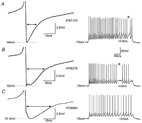
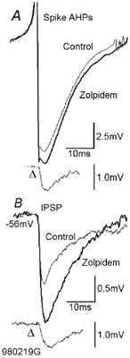
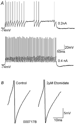
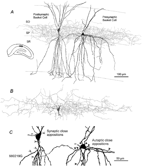
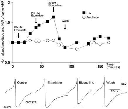
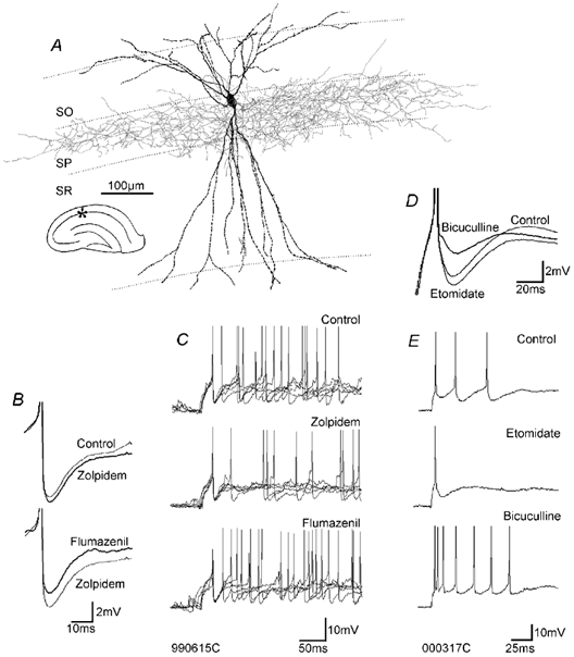
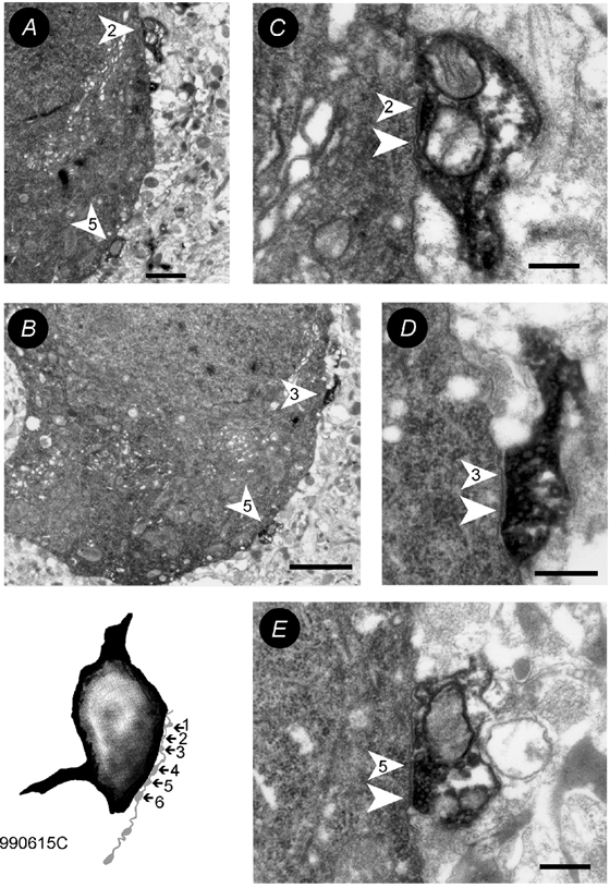
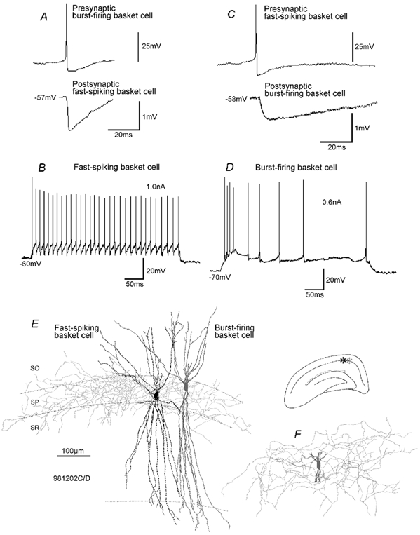
To determine whether autaptic inhibition plays a functional role in the adult hippocampus, the action potential afterhyperpolarisations (spike AHPs) of CA1 interneurones were investigated in 25 basket, three bistratified and eight axo-axonic cells. The spike AHPs showed two minima in all regular-spiking (5), burst-firing (3) and in many fast-spiking cells (17:28). The fast component had a time-to-peak (TTP) of 1.2 +/- 0.5 ms, the slower TTP was very variable (range of 3.3-103 ms). The AHP width at half-amplitude (HW) was 12.5 +/- 5.7 ms in fast-spiking, 29.3 +/- 18 ms in regular-spiking and 99.7 +/- 42 ms in burst-firing cells. Axo-axonic cells never establish autapses, and the fast-spiking variety showed narrow (HW: 3.9 +/- 0.7 ms) spike AHPs with only one AHP minimum (TTP: 0.9 +/- 0.1 ms). When challenged with GABA(A) receptor modulators, spike AHPs in basket and bistratified cells were enhanced by zolpidem (HW by 18.4 +/- 6.2 % in 10:15 cells tested), diazepam (45.2 +/- 0.5 %, 6:7), etomidate (43.9 +/- 36 %, 6:8) and pentobarbitone sodium (41 %, 1:1), and were depressed by bicuculline (-41 +/- 5.7 %, 5:8) and picrotoxin (-54 %, 1:1), and the enhancement produced by zolpidem was reduced by flumazenil (-31 +/- 13 %, relative to the AHP HW during exposure to zolpidem, 3:4). Neuronal excitability was modulated in parallel. The spike AHPs of three axo-axonic cells tested showed no sensitivity to etomidate, pentobarbitone or diazepam. Interneurone-to-interneurone inhibitory postsynaptic potentials (IPSPs), studied with dual intracellular recordings, had time courses resembling those of the spike AHPs. The IPSP HW was 13.4 +/- 2.8 ms in fast-spiking (n = 16) and 28.7 +/- 5.8 ms in regular-spiking/burst-firing cells (n = 6), and the benzodiazepine1-selective modulator zolpidem strongly enhanced these IPSPs (45 +/- 28 %, n = 5). Interneurones with spike AHPs affected by the GABA(A) receptor ligands exhibited 3.8 +/- 1.9 close autaptic appositions. In three basket cells studied at the ultrastructural level 6 of 6, 1 of 2 and 1 of 2 close appositions were confirmed as autapses. Therefore, in the hippocampus autaptic connections contribute to spike AHPs in many interneurones. These autapses influence neuronal firing and responses to GABA(A) receptor ligands.


Figures

Figure 1
Figure 1. Variability of spike AHPs in fast-spiking basket cells
The shape of the AHPs (left panel) in individual cells varied from triangular (A) via tri-phasic (B) to rounded (C). In these examples spiking was elicited with current injections of 0.6, 0.2 and 0.3 nA, and AHPs following the first, second and second spike, respectively, were averaged. However, for the analysis of drug effects (e.g. Figs 2-5) only spike AHPs following the first spike were analysed. The double arrows indicate the AHP width at half-amplitude. The resting membrane potential in all three cells was −70 mV. The action potentials are truncated. The right panel shows examples of the discharge pattern of the individual neurones. *A prominent feature in many fast-spiking cells is the interrupted discharge in the gamma frequency range (A and B).
Figure 2
Figure 2. Modulation of spike AHPs and IPSPs by zolpidem
A, the spike AHP of a fast-spiking presynaptic basket cell was increased by 0.2 µm zolpidem (average from 15–30 min of drug application). The traces indicated by Δ show the difference between control and drug application traces. The IPSPs generated by this basket cell in another postsynaptic fast-spiking basket cell were increased in parallel (B). As autaptic IPSPs superimpose on spike AHPs, they will be distorted by the rapid membrane potential changes associated with the AHP. These difference traces do not therefore indicate the shape or time course of the event that would have been recorded in isolation. For the anatomy of the pre- and postsynaptic cell see Fig. 6.
Figure 5
Figure 5. The spike AHP of a fast-spiking axo-axonic cell was not enhanced by etomidate
A, current injection of intermediate intensities (e.g. 0.2 nA) into the axo-axonic cell elicited interrupted fast-spiking discharge. Injection of higher current intensities (e.g. 0.4 nA) elicited uninterrupted spiking. B, spike AHPs of fast-spiking axo-axonic cells were monophasic and very brief. This spike AHP was not affected by etomidate.
Figure 6
Figure 6. Light microscopic reconstruction of the pair of synaptically connected basket cells shown in Fig. 2
A, the presynaptic basket cell (black soma-dendrites and light grey axonal arbour) generated IPSPs in the postsynaptic basket cell (dark grey, soma-dendrites only). B, axonal arbour of the postsynaptic basket cell. C, soma and proximal dendrites of the pre- and postsynaptic cell. The arrowheads indicate the sites of close apposition of the presynaptic cell's axon with the postsynaptic cell (left, semi-filled arrowheads) and with its own somato-dendritic domain (right, filled arrowheads). This magnification reveals the sparsely spiny dendrites of the presynaptic and the smooth dendrites of the postsynaptic cell. Abbreviations: ax = axon-initial segment, SO = stratum oriens, SP = stratum pyramidale, SR = stratum radiatum.
Figure 4
Figure 4. Modulation of the spike AHP by etomidate and bicuculline
On a normalised scale, the AHP amplitude (○) and the AHP width at half-amplitude (HW, ▪) are plotted against time. The arrows indicate the onset of drug applications. The lower part shows spike AHPs from different phases of the experiment.
Figure 3
Figure 3. GABAA receptor ligands modulate the spike AHPs and discharge of fast-spiking and regular-spiking interneurones
A, light microscopic reconstruction of a fast-spiking basket cell (the ultrastructure of its autapses is shown in Fig. 7). B, spike AHPs of the fast-spiking cell were enhanced by zolpidem and reversed by flumazenil, even beyond control levels. C, the modulation of spike AHPs by zolpidem and flumazenil was accompanied by reduced and increased neuronal excitability, respectively. In each panel, five individual traces are superimposed, with the first spikes superimposed. D, the spike AHPs of this regular-spiking basket cell were increased by etomidate and decreased by bicuculline. E, the increase in the spike AHP was accompanied by a reduction in neuronal excitability and vice versa. The discharge in C and E was elicited by injection of current pulses of 0.4 nA and 0.15 nA, respectively. Abbreviations: SO = stratum oriens, SP = stratum pyramidale, SR = stratum radiatum.
Figure 7
Figure 7. Electron microscopic verification of autapses
The inset shows the drawing tube reconstruction of the soma (black) and a part of the axon (light grey) of the fast-spiking basket cell shown in Fig. 3A. The six close appositions were all confirmed to be synaptic contacts. The electron microscopic images of contacts 1 (A,C), 3 (B,D) and 5 (B,E) are shown at low (A,B) and high (C-E) magnification. The arrowheads point towards the postsynaptic densities, the numbers correspond to the number of close appositions observed at the light microscopic level. Scale bars: A = 1µm, B = 2µm, C-E = 250 nm.
Figure 8
Figure 8. Properties of IPSPs in a pair of reciprocally connected basket cells (one fast-spiking cell and one burst-firing cell)
A, spikes in the burst-firing basket cell generated IPSPs with a fast time course in the postsynaptic fast-spiking cell. B, discharge characteristics of the fast-spiking cell. Current injection as indicated. C, spikes in the fast-spiking basket cell generated IPSPs in the burst-firing basket cell with a relatively slow time course. D, discharge characteristics of the burst-firing basket cell. E and F, light microscopic reconstruction of the fast-spiking and the burst-firing basket cell. E, soma/dendrites (black) and axonal arbour (light grey) of the fast-spiking cell and the soma/dendrites (dark grey) of the burst-firing cell are shown. F, cropped soma/dendrites (dark grey) and axonal arbour (light grey) of the burst-firing cell.

Similar articles

Cited by

References

    1. Ali AB, Bannister AP, Thomson AM. IPSPs elicited in CA1 pyramidal cells by putative basket cells in slices of adult rat hippocampus. Eur J Neurosci. 1999;11:1741–1753. - PubMed
    1. Aoki T, Baraban SC. Properties of a calcium-activated K+ current on interneurons in the developing rat hippocampus. J Neurophysiol. 2000;83:3453–3461. - PubMed
    1. Bekkers JM, Stevens CF. Excitatory and inhibitory autaptic currents in isolated hippocampal neurons maintained in cell culture. Proc Natl Acad Sci U S A. 1991;88:7834–7838. - PMC - PubMed
    1. Buhl EH, Han Z-S, Lörinczi Z, Stezhka VV, Karnup SV, Somogyi P. Physiological properties of anatomically identified axo-axonic cells in the rat hippocampus. J Neurophysiol. 1994;71:1289–1307. - PubMed
    1. Chen G, Van Den Pol AN. Multiple NPY receptors coexist in pre- and postsynaptic sites: inhibition of GABA release in isolated self-innervating SCN neurons. J Neurosci. 1996;16:7711–7724. - PMC - PubMed

Publication types

MeSH terms

LinkOut - more resources