. 2003 Feb 1;546(Pt 3):823-36.
doi: 10.1113/jphysiol.2002.030775.

Essential role of rho kinase in the Ca2+ sensitization of prostaglandin F(2alpha)-induced contraction of rabbit aortae

Affiliations

Essential role of rho kinase in the Ca2+ sensitization of prostaglandin F(2alpha)-induced contraction of rabbit aortae

Katsuaki Ito et al. J Physiol. .

Abstract

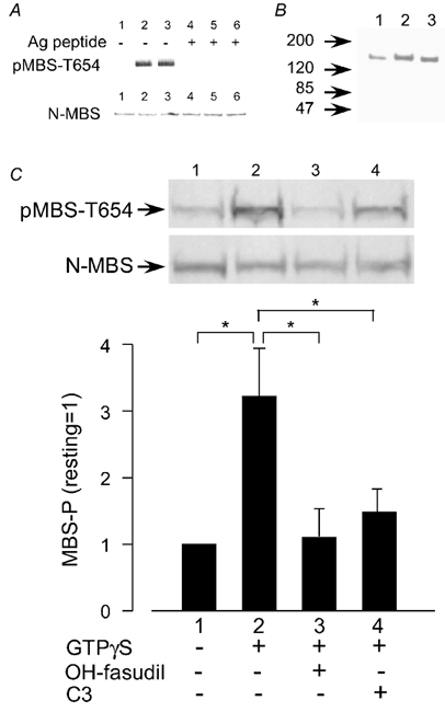
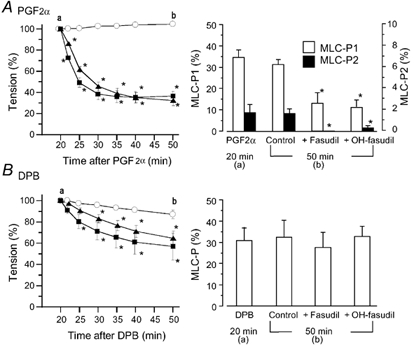
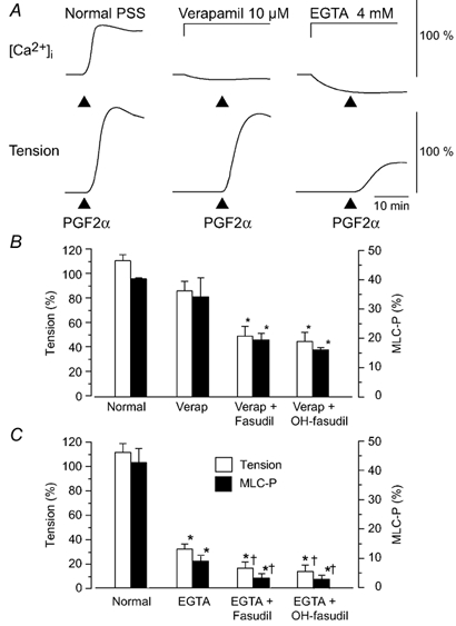
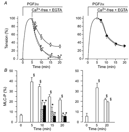
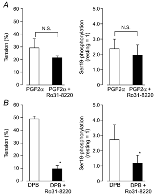
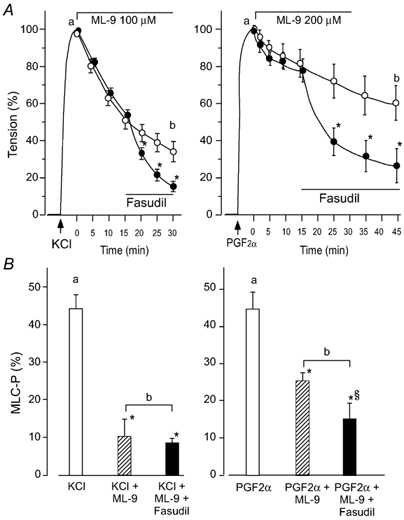
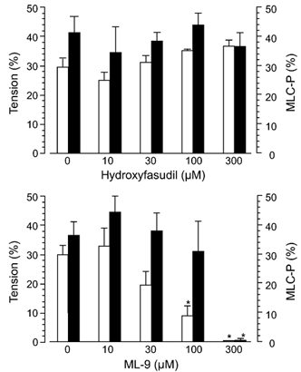
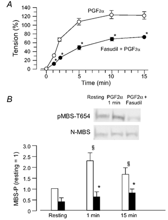
Inhibition of dephosphorylation of the 20 kDa myosin light chain (MLC(20)) is an important mechanism for the Ca(2+)-induced sensitization of vascular smooth muscle contraction. We investigated whether this mechanism operates in prostaglandin F(2alpha) (PGF(2alpha))-induced contraction of rabbit aortic smooth muscle and, if so, whether protein kinase C (PKC) or rho-associated kinase (rho kinase) contribute to the inhibition of dephosphorylation. In normal medium, PGF(2alpha) (10 microM) increased the phosphorylation of MLC(20) and developed tension. The rho-kinase inhibitors fasudil and hydroxyfasudil inhibited these changes, despite having no effect on a phorbol-ester-induced MLC(20) phosphorylation. After treatment with verapamil or chelation of external Ca(2+) with EGTA, PGF(2alpha) increased the MLC(20) phosphorylation and tension without an increase in [Ca(2+)](i), all of which were sensitive to fasudil and hydroxyfasudil. ML-9, a MLC kinase inhibitor, quickly reversed the KCl-induced MLC(20) phosphorylation and contraction to the resting level. However, fractions of PGF(2alpha)-induced contraction and MLC(20) phosphorylation were resistant to ML-9 but were sensitive to fasudil. Ro31-8220 (10 microM), a PKC inhibitor, did not affect the phosphorylation of MLC(20) and the tension caused by PGF(2alpha), thus excluding the possibility of the involvement of PKC in the PGF(2alpha)-induced MLC(20) phosphorylation. PGF(2alpha) increased phosphorylation at Thr654 of the myosin binding subunit (MBS) of myosin phosphatase, which is a target of rho kinase, and fasudil decreased the phosphorylation. These data suggest that the PGF(2alpha)-induced contraction is accompanied by the inhibition of MLC(20) dephosphorylation through rho kinase-induced MBS phosphorylation, leading to Ca(2+) sensitization of contraction. An actin-associated mechanism may also be involved in the PGF(2alpha)-induced sensitization.


Figures

Figure 7
Figure 7. Characterization of the antibody pMBS-T654
A, detection of rho kinase-induced phosphorylation of the myosin binding subunit (MBS) by pMBS-T654 in vitro. Recombinant MBS was phosphorylated by rho kinase for 0 min (lanes 1 and 4), 10 min (lanes 2 and 5) or 20 min (lanes 3 and 6) and subjected to SDS-PAGE, followed by Western blotting with pMBS-T654 (upper panel) or with the antibody N-MBS, which recognizes the N-terminus of MBS (lower panel), in the absence (lanes 1–3) or presence (lanes 4–6) of phosphorylated antigen peptide (T654-phosphopeptide). B, PGF-induced phosphorylation at Thr654 of MBS in the rabbit carotid artery. Extracts of arteries before stimulation (lane 1) or 1 (lane 2) and 2 min (lane 3) after the stimulation with PGF (30 µm) were subjected to SDS-PAGE followed by Western blotting with pMBS-T654. C, GTPγS-induced phosphorylation at Thr654 of MBS (MBS-P) and its inhibition by hydroxyfasudil or C3 toxin in permeabilized pig aortic cells. Cells were treated with GTPγS for 10 min. When used, hydroxyfasudil (OH-fasudil, 10 µm) or C3 toxin (1 µg ml−1) was used to pretreat cells for 15 min before application of GTPγS. Photographs: band reacted with pMBS-T654 (upper panel) or N-MBS (lower panel). MBS phosphorylation is expressed as a percentage of the level in the resting state. Data are means ± s.e.m. (n = 4). *P < 0.05.
Figure 1
Figure 1. Effects of fasudil and hydroxyfasudil on the prostaglandin F (PGF)- or 12-deoxyphorbol 13-isobutyrate (DPB)-induced contraction and 20 kDa myosin light chain (MLC20) phosphorylation in rabbit aortae
Fasudil (▪) or hydroxyfasudil (OH-fasudil, (Δ) at 10 µm was added 20 min after the application of 10 µm PGF (A) or 1 µm DPB (B). Left panels, a change in tension after the addition of fasudil or hydroxyfasudil (○, agonist alone). Tension is expressed as a percentage of contraction just before the addition of fasudil or hydroxyfasudil. In the right panels, MLC20 phosphorylation (MLC-P) was observed 20 min after the addition of PGF or DPB (just before the addition of fasudil or hydroxyfasudil (a) or 50 min after the addition of PGF or DPB (b), and is expressed as a percentage of phosphorylated MLC20 of total MLC20. MLC20 phosphorylation in the resting state was 8–9 % in each case (data not shown). Since PGF caused diphosphorylation of MLC20, monophosphorylation (MLC-P1, open columns) and diphosphorylation (MLC-P2, black columns) are shown (A, right panel). Data are presented as the mean ± s.e.m. (n = 5–6). * Significantly different from the time-matched control (P < 0.05).
Figure 2
Figure 2. Changes in[Ca2+]i, tension and MLC20 phosphorylation due to PGF under normal conditions or conditions where an increase in [Ca2+]i was inhibited
A, examples of changes in[Ca2+]i in normal physiological saline solution (PSS), in the presence of verapamil (10 µm) or EGTA (4 mm). For[Ca2+]i and tension, 100 % represents the change induced by 65.4 mm KCl, which was obtained before the application of PGF. Verapamil or EGTA was applied 10 min before the addition of PGF (10 µm). B, summarized data of the PGF-induced contraction and MLC20 phosphorylation in the normal medium or in the presence of verapamil and fasudil or hydroxyfasudil (OH-fasudil). Developed tension is expressed as a percentage of the maximum contraction to 65.4 mm KCl. C, summarized data of the PGF-induced contraction and MLC20 phosphorylation in the normal medium or in the presence of EGTA and fasudil or OH-fasudil. In B and C, MLC20 phosphorylation (MLC-P) was measured at 5 min after the addition of PGF (10 µm). When used, fasudil (10 µm) or hydroxyfasudil (OH-fasudil, 10 µm) was added 30 min before PGF. The protocol in B and C was the same as that shown in A, but the experiments were performed in a different series. MLC20 phosphorylation under the resting condition in normal PSS was 8.9 ± 2.4 % (data not shown). Verap: verapamil.
Figure 3
Figure 3. Effects of fasudil, hydroxyfasudil or ONO-RS-082 on MLC20 phosphorylation and tension maintained in Ca2+-free, EGTA solution in the presence of PGF
A, change in tension. After PGF (10 µm)-induced contraction had reached a peak, the external medium was changed from normal PSS to Ca2+ free, 1 mm EGTA solution (5 min in the figure; ○, PGF alone). When used, fasudil (δ, 10 µm) or hydroxyfasudil (□, 10 µm) was added at the time when the external Ca2+ was omitted (the left panel). ONO-RS-082 (•, 5 µm, the right panel) was applied 15 min before the addition of PGF. Data are expressed as a percentage of the maximal contraction to PGF in normal medium (n = 6-7). B, MLC20 phosphorylation (MLC-P) measured in the resting state (0 min), just before switching the medium to Ca2+ free, EGTA solution (5 min), 5, 10 and 15 min after Ca2+ removal (10, 15 and 20 min after the addition of PGF). Open columns: controls; hatched columns: fasudil-treated muscles; filled columns: hydroxyfasudil-treated muscles; grey column: ONO-RS-082-treated muscles. * Significantly different from the level in the absence of fasudil or hydroxyfasudil (P < 0.05); § significantly different from the resting level (P < 0.05).
Figure 4
Figure 4. Effects of the protein kinase C (PKC) inhibitor Ro31-8220 on phosphorylation of MLC20 at Ser19 and contraction induced by PGF and DPB
A, the effects of Ro31-8220 on the PGF-induced inhibition of Ser19 dephosphorylation and relaxation. The protocol was the same as in Fig. 3. When the external medium was changed from normal PSS to Ca2+-free, 1 mm EGTA solution, Ro31-8220 (5 µm) was simultaneously added to one group (filled columns) but not to the control group (open columns). B, the effects on the DPB-induced inhibition of Ser19 dephosphorylation and relaxation. After 65.4 mm KCl caused a maximal contraction (5 min), the external medium was switched to Ca2+ free, 1 mm EGTA solution (5.4 mm K+) containing 1 µm DPB. When present, Ro31-8220 (5 µm) was added simultaneously with DPB. Data were taken 15 min after the omission of external Ca2+. The left panels represent a relative value of the 65.4 mm KCl-induced contraction, which was obtained before PGF. The right panels are Ser19 phosphorylation expressed as a value relative to the phosphorylation in the resting state. Data are mean ± s.e.m. (n = 4). * Significantly different from DPB alone (P < 0.05, N.S. not significant).
Figure 5
Figure 5. Sensitivity of KCl- and PGF-induced contraction and MLC20 phosphorylation to ML-9 and fasudil
After KCl (65.4 mm, left)- or PGF (10 µm, right)-induced contraction reached a respective peak (5 min), ML-9 (100 or 200 µm, respectively) was applied. Fasudil (10 µm) was added 15 min later. A, a change in tension in fasudil-treated (•) or untreated muscles (○). The ordinate represents the relative tension of maximum contraction to KCl or PGF, respectively. B, MLC20 phosphorylation (MLC-P) measured in the protocol shown in A. Data were sampled just before the addition of ML-9 (open columns, corresponding to a in A) and 30 min after the addition of ML-9 for KCl-induced contraction or 45 min after addition of ML-9 for PGF-induced contraction (corresponding to b in A). Hatched columns: in the absence of fasudil, filled columns: in the presence of fasudil. * Significantly different from the level before the addition of ML-9 (P < 0.05), § significantly different from the level in the absence of fasudil (P < 0.05n = 5–8).
Figure 6
Figure 6. Differential effects of hydroxyfasudil and ML-9 on the calyculin A-induced contraction and MLC20 phosphorylation
Open columns: tension expressed as a percentage of the maximum contraction due to 65.4 mm KCl, which was obtained before the addition of calyculin A, filled columns: MLC-P. At 30 min after the application of calyculin A (300 nm), hydroxyfasudil or ML-9 at each concentration was added. * Significantly different from the level in the absence of ML-9 (P < 0.05).
Figure 8
Figure 8. PGF-induced phosphorylation of the MBS of phosphatase, and its inhibition by fasudil
A, time-course of the PGF (10 µm)-induced contraction in the absence (○) or presence (•) of fasudil (10 µm). When present, fasudil was applied 15 min before addition of PGF. B, (photographs) example of Western blots with the pMBS-T654 antibody or the N-MBS antibody. Samples were taken before the addition of PGF (resting) and 1 min after PGF. Bar graph, quantified phosphorylation of MBS (MBS-P) in the resting state, 1 or 15 min after the addition of PGF in the absence (open columns) or presence (filled columns) of fasudil. The ratio of the density of a band with the pMBS-T654 antibody to that with the N-MBS antibody in the resting state is referred as 1. Data are presented as the means ± s.e.m. of four experiments. * Significantly different from the resting level (P < 0.05); § significantly different from PGF alone (P < 0.05).

Similar articles

Cited by

References

    1. Balwierczak JL. The relationship of KCl- and prostaglandin F2α-mediated increases in tension of the porcine coronary artery with changes in intracellular Ca2+ measured with fura-2. Br J Pharmacol. 1991;104:373–378. - PMC - PubMed
    1. Beavo JA, Bechtel PJ, Krebs EG. Preparation of homogeneous cyclic AMP-dependent protein kinase(s) and its subunits from rabbit skeletal muscle. Methods Enzymol. 1974;38:299–308. - PubMed
    1. Bradley AL, Morgan KG. Alteration in cytoplasmic calcium sensitivity during porcine coronary artery contraction as detected by aequorin. J Physiol. 1987;385:437–448. - PMC - PubMed
    1. Buus CL, Aalkjar C, Nilsson H, Juul B, Moller JV, Mulvany MJ. Mechanisms of Ca2+ sensitization of force production by noradrenaline in rat mesenteric small arteries. J Physiol. 1998;510:577–590. - PMC - PubMed
    1. Chehrazi BB, Giri S, Joy RM. Prostaglandins and vasoactive amines in cerebral vasospasm after aneurysmal subarachnoid hemorrhage. Stroke. 1989;20:217–224. - PubMed

MeSH terms

LinkOut - more resources