. 2003 Feb 1;546(Pt 3):859-67.
doi: 10.1113/jphysiol.2002.031369.

PSD-95 regulates synaptic transmission and plasticity in rat cerebral cortex

Affiliations

PSD-95 regulates synaptic transmission and plasticity in rat cerebral cortex

Jean-Claude Béïque et al. J Physiol. .

Abstract

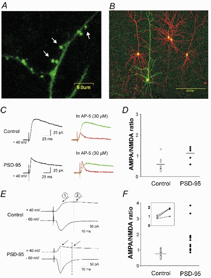
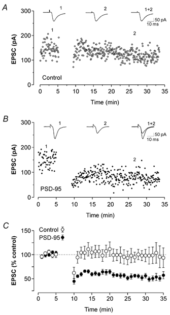
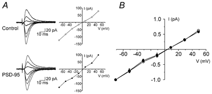
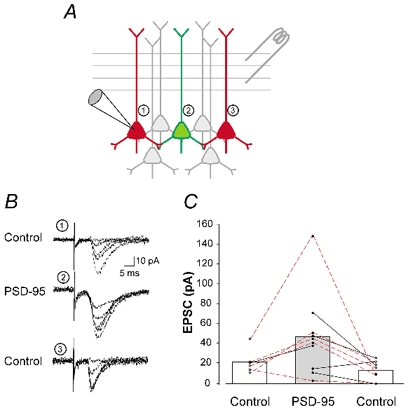
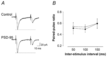
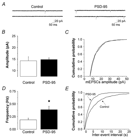
PSD-95 is one of the most abundant proteins found in the postsynaptic density of excitatory synapses. However, the precise functional role played by PSD-95 in regulating synaptic transmission and plasticity remains undefined. To address this issue, we have overexpressed PSD-95 in cortical pyramidal neurons in organotypic brain slices using particle-mediated gene transfer and assessed the consequences on synaptic transmission and plasticity. The AMPA receptor/NMDA receptor (AMPAR/NMDAR) ratio of evoked EPSCs recorded at +40 mV was greater in PSD-95-transfected pyramidal neurons than in controls. This difference could not be accounted for by a change in rectification of AMPAR-mediated synaptic currents since the current-voltage curves obtained in controls and in PSD-95-transfected neurons were indistinguishable. However, the amplitude of AMPAR-mediated evoked EPSCs was larger in PSD-95-transfected neurons compared to matched controls. Paired-pulse ratio analysis suggested that overexpression of PSD-95 did not alter presynaptic release probability. Transfection of PSD-95 was further accompanied by an increase in the frequency, but not amplitude, of AMPAR-mediated mEPSCs. Together, these results indicate that transfection of PSD-95 increased AMPAR-mediated synaptic transmission. Furthermore, they suggest that this phenomenon reflects an increased number of synapses expressing AMPARs rather than an increased number or function of these receptors at individual synapses. We tested the consequences of these changes on synaptic plasticity and found that PSD-95 transfection greatly enhanced the probability of observing long-term depression. These results thus identify a physiological role for PSD-95 and demonstrate that this protein can play a decisive role in controlling synaptic strength and activity-dependent synaptic plasticity.


Figures

Figure 1
Figure 1. Expression of PSD-95 in pyramidal neurons of the prefrontal cortex increases the AMPAR/NMDAR ratio
A, confocal image taken after 2 days in vitro illustrating the preferential targeting of PSD-95-GFP fusion protein to dendritic spines (see arrows). B, illustration of the experimental preparation used for these experiments. Prefrontal cortex pyramidal neurons in this slice were transfected with PSD-95/EGFP (green) and (in some cases) with pcDNA3.1/DsRed (red) to serve as within-slice transfected controls. C, current traces recorded at +40 mV from neurons transfected with pcDNA3.1/EGFP (control) or PSD95/EGFP, before (black trace) and after (red trace) administration of 30 µm (d)-AP-5. The green traces depict the NMDAR (AP-5-sensitive) component of the evoked current obtained by subtraction. D, computed AMPAR/NMDAR ratios obtained from PSD-95-transfected neurons (n = 6) and controls (5 cells transfected with pcDNA3.1/EGFP and 3 untransfected neurons; P < 0.02). E, current traces recorded at −60 and +40 mV from a control (pcDNA3.1/EGFP) and from a PSD-95-transfected neuron. By superimposing the traces, the AMPAR (1) and NMDAR (2) components of the traces at +40 mV were estimated. F, computed AMPAR/NMDAR ratios obtained using this method for PSD-95-transfected (n = 14) and control neurons (6 untransfected cells and 7 cells transfected with empty plasmid (i.e. pcDNA3.1/EGFP; sister slice comparisons, n = 5; or pcDNA3.1/DsRed; same slice comparisons, n = 2). A subset of these experiments involving pairwise sequential recordings from neighbouring neurons is re-plotted in the inset. PSD-95 transfection increased the AMPAR/NMDAR ratio (P < 0.01 for pooled data and P < 0.03 for pairwise recordings).
Figure 6
Figure 6. PSD-95 overexpression favours the emergence of LTD
A, in this untransfected control neuron, pairing presynaptic stimulation and postsynaptic depolarization did not induce any significant change in synaptic strength. Each data point represents the amplitude of EPSCs recorded at −70 mV. B, in a PSD-95-transfected cell, the same pairing protocol resulted in the appearance of robust LTD. C, ensemble average plot for the experiments illustrated in panels A and B. The pairing protocol did not induce any significant changes in EPSC amplitudes in control cells (○; n = 10; 5 untransfected cells recorded in a sequential pairwise fashion with a PSD-95-transfected neurons, 3 cells transfected with pcDNA3.1/EGFP and 2 untransfected cells). In contrast, the same pairing protocol induced a significant and lasting reduction of EPSCs amplitude in PSD-95-transfected neurons (•; n = 12; P < 0.01).
Figure 2
Figure 2. PSD-95 does not change the rectification properties of AMPAR-mediated synaptic currents
A, current traces and I-V relationships for AMPAR-mediated EPSCs in a control (non-transfected) and a PSD-95-transfected cell. B, average I-V relationships for AMPAR-mediated EPSCs in PSD-95-transfected (n = 5) and control neurons (n = 7; 6 untransfected neurons and 1 neuron transfected with pcDNA3.1/EGFP). The experiments were carried out in the presence of 30 µm (d)-AP-5. Data were normalized for every cell to the amplitude of the synaptic current obtained at −71 mV.
Figure 3
Figure 3. PSD-95 increases the amplitude of AMPAR-mediated EPSCs
A, illustration of the experimental approach used for the experiments depicted in B and C. Evoked AMPAR-mediated EPSCs recorded from a PSD-95-transfected neuron (green) were compared to those recorded from 2 control neurons (red), spatially bracketing the transfected cell. B, superimposed individual current traces from 3 neurons in one of the triads illustrated in C. C, amplitude of EPSCs recorded from control (n = 13 untransfected and n = 2 pcDNA3.1/DsRed-transfected) and PSD-95-transfected neurons (n = 10). The data are plotted as a graph (control-PSD-95-control) so as to illustrate the spatial organization in which the recordings were obtained (as depicted in A). Triad recordings (n = 5) are outlined by the red connecting lines. In some cases, only paired recordings could be obtained (n = 5; black lines). PSD-95-transfection resulted in a significant increase in the amplitude of AMPAR-mediated evoked EPSCs (P < 0.05 for triads and P < 0.02 for the pooled data (triad and pairwise recordings).
Figure 4
Figure 4. PSD-95 does not alter presynaptic release probability
A, current traces from a control (pcDNA3.1/EGFP) and from a PSD-95-transfected neuron illustrating the response to two successive stimulations (50 ms interval). B, paired-pulse ratios calculated at three distinct inter-stimulus intervals (ISI) were not different between controls (n = 4; 3 cells transfected with pcDNA3.1/EGFP and 1 untransfected neuron) and PSD-95-transfected neurons (n = 4; P > 0.5 for all intervals).
Figure 5
Figure 5. PSD-95 increases the frequency, but not the amplitude of AMPAR mEPSCs
A, current traces (recorded in the presence of TTX, bicuculline and (d)-AP-5) from a control (untransfected) cell and a PSD-95-transfected cell. B, PSD-95 (n = 6) did not induce any significant change in the amplitude of mEPSC (n = 8 untransfected control). C, average cumulative probability plot for mEPSC amplitudes in all PSD-95-transfected and control neurons. D, PSD-95 resulted in a significant increase in the frequency of mEPSCs (P < 0.02). E, average cumulative probability plots for the mEPSC inter-event intervals in all PSD-95-transfected and control neurons.

Similar articles

Cited by

References

    1. Craven SE, Bredt DS. PDZ proteins organize synaptic signaling pathways. Cell. 1998;93:495–498. - PubMed
    1. Edmonds B, Gibb AJ, Colquhoun D. Mechanisms of activation of glutamate receptors and the time course of excitatory synaptic currents. Annu Rev Physiol. 1995;57:495–519. - PubMed
    1. El Husseini AE, Schnell E, Chetkovich DM, Nicoll RA, Bredt DS. PSD-95 involvement in maturation of excitatory synapses. Science. 2000;290:1364–1368. - PubMed
    1. El Husseini AE, Schnell E, Dakoji S, Sweeney N, Zhou Q, Prange O, Gauthier-Campbell C, Aguilera-Moreno A, Nicoll RA, Bredt DS. Synaptic strength regulated by palmitate cycling on PSD-95. Cell. 2002;108:849–863. - PubMed
    1. Haj-Dahmane S, Andrade R. Muscarinic activation of a voltage-dependent cation nonselective current in rat association cortex. J Neurosci. 1996;16:3848–3861. - PMC - PubMed

Publication types

MeSH terms

Substances

LinkOut - more resources