. 2003 Feb 3;197(3):333-41.
doi: 10.1084/jem.20021639.

Extrathymic T cell lymphopoiesis: ontogeny and contribution to gut intraepithelial lymphocytes in athymic and euthymic mice

Affiliations

Extrathymic T cell lymphopoiesis: ontogeny and contribution to gut intraepithelial lymphocytes in athymic and euthymic mice

Delphine Guy-Grand et al. J Exp Med. .

Abstract

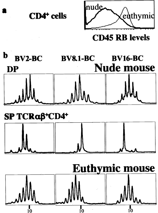
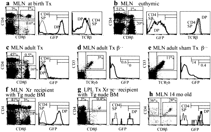
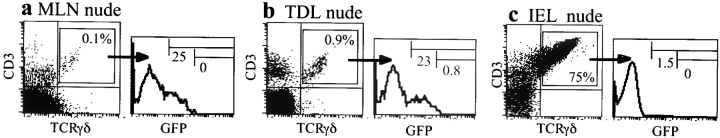
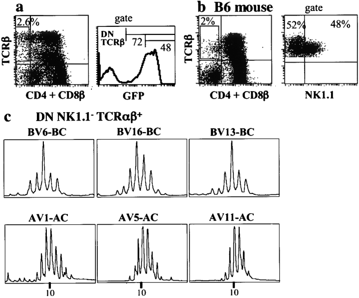
In the absence of thymopoiesis, T lymphocytes are nevertheless present, mainly in the gut epithelium. Ontogeny of the extrathymic pathway and the extent of its involvement in euthymic mice are controversial. These questions have been addressed by assessing the expression of recombinase activating gene (RAG) through the use of green fluorescent protein RAG2 transgenic mouse models. In athymic mice, T lymphopoiesis occurs mainly in the mesenteric lymph node and less in the Peyer's patches. Ontogenic steps of this lymphopoiesis resemble those of thymopoiesis, but with an apparent bias toward gamma delta T cell production and with a paucity of oligoclonal alpha beta T cells possibly resulting from a deficit in positive selection. Whether in athymic or euthymic mice, neither T intraepithelial lymphocytes (IEL) nor cryptopatch cells (reported to contain precursors of IEL) displayed fluorescence indicating recent RAG protein synthesis. Newly made T cells migrate from the mesenteric node into the thoracic duct lymph to reach the gut mucosa. In euthymic mice, this extrathymic pathway is totally repressed, except in conditions of severe lymphocytic depletion. Thus, in normal animals, all gut T IEL, including CD8 alpha alpha(+) cells, are of thymic origin, CD8 alpha alpha(+) TCR alpha beta(+) IEL being the likely progeny of double negative NK1-1(-) thymocytes, which show polyclonal V alpha and V beta repertoires.


Figures

Figure 1.
Figure 1.
GFPH lymphocytes in tissue sections from nude or euthymic mice. (a) MLN, nude mouse: clusters of fluorescent GFPH cells located in some medullary cords (empty spaces are lymphatic vessels of the medulla). (b) PP, nude mouse: rare GFPH cells located in an interfollicular T area near lymphatic vessels. (c) Cryptopatch, nude mouse: lack of GFPH cells in the patch and adjacent epithelium (∼10 cryptopatches were explored in various mice). (d) MLN, euthymic mouse: absence of GFPH cells in the medulla. ×70.
Figure 2.
Figure 2.
Analysis of GFP expression during T lymphopoiesis in athymic mice and thymopoiesis. (a–d) MLN (a and b) and thymus (c and d). (a and c) Comparative features of DP and SP cells analyzed for GFP (middle histograms) and TCRβ (right histograms) expressions. Note that DP cells express less TCRβ chains than SP and that there is a small difference between thymus and MLN DP cells in this respect. (b and d) TN CD25H cells (after gating out CD3+, CD4+, CD8+, and CD19+ cells) analyzed for GFP content of CD44+ DN2 (middle histograms) and CD44 DN3 cells (right histograms). All other TN cells were GFP and most of the MLN CD25L cells had the phenotype of cryptopatch cells (reference 8). Lack or rarity of DN4 cells (CD44 CD25) results from gating out CD3 and coreceptors bearing cells with potent antibodies binding even cells with trace amounts of these molecules, as is the case of most DN4 cells. The percentages of c-Kit+ and IL7-Rα+ in CD25+ TN MLN cells and thymocytes are shown in the right panels. (e) Thymus: γδ+ cells analyzed for GFP content. (f–h) LP lymphocytes analyzed for GFP content. (f) DP cells are GFPH and (g) TN CD25H cells (after gating out of cells as described above) are GFPH. (h) TN c-Kit+ CD25+/− cells, belonging to a population of CD44+ Thy-1+/− IL7-R+ CD4+/− cells (not depicted), i.e., with the phenotype of cryptopatch cells (references and 20), are GFP.
Figure 3.
Figure 3.
Exploration of the memory phenotype and of the TCRβ repertoire of MLN αβ+ CD4+ cells of nude and euthymic mice. (a) MLN CD4+ cells from a nude mouse express the phenotype of memory cells, in contrast with comparable cells from an euthymic mouse. Cells were triply labeled with anti-TCRβ, anti-CD4, and anti-CD45RB mAbs. (b) The TCRβ repertoire of MLN DP cells from a nude mouse shows the gaussian-like profile characteristic of polyclonal populations (top), whereas that of SP CD4+ cells is oligoclonal (middle), in contrast with that of comparable cells from an euthymic mouse (bottom).
Figure 4.
Figure 4.
Analysis of lymphopoiesis in various Tx or euthymic mice. (a) MLN cells, mouse Tx at birth (compare with Fig. 2 a). (b) MLN cells, 7-wk-old euthymic mouse, sham Tx (match of mouse with adult Tx shown in c). Presence of some DP with GFP and TCRβ expression similar to that of SP CD4+ cells (right panels). Note that many CD4+ cells are GFPL. (c) MLN, 7-wk-old mouse 2 wk after thymectomy. All DP and SP cells are GFP (compare with b). (d and e) MLN cells from 8-wk-old Tg TCRβ−/− mice, Tx (d) or sham Tx (e) 2 wk earlier, used for easy study of γδ+ cells. GFPL cells are absent in the Tx mouse. (f) MLN cells of lethally irradiated non-Tg mouse reconstituted 18 d earlier with bone marrow cells from a nude Tg mouse. DP cells are GFPH. Note that MLN cell recovery was 10-fold less than that from normal mice. (g) Lymphocytes isolated from the LP of a Tx RAGγc−/− mutant mouse reconstituted 36 d earlier with nude Tg mouse bone marrow cells (note that some CD4+ GFP cells, originating from the graft, are detectable). (h) MLN cells from a 14-mo-old mouse (cell recovery was ∼20-fold less than in younger mice). DP cells with GFP expression comparable to that of DP thymocytes.
Figure 5.
Figure 5.
GFPL γδ1 T cells in nude mice; comparison between MLN, circulating cells, and IEL. γδ+ T from MLN (a), thoracic duct lymphocytes (TDL; b), and IEL (c) analyzed for GFP expression.
Figure 6.
Figure 6.
Analyses of DN αβ1 thymocytes. Studies of (a) GFP content and (b) NK1-1 expression (in this last case, cells were from a B6 mouse because the Tg mice do not express NK1.1). (c) TCRα and β repertoires of DN NK1.1 thymocytes: polyclonal repertoires expressing chains different from that peculiar to NKT cells.

References

    1. Park, S.H., D. Guy-Grand, F.A. Lemonnier, C.R. Wang, A. Bendelac, and B. Jabri. 1999. Selection and expansion of CD8αα+ T cell receptor αβ+ intestinal intraepithelial lymphocytes in the absence of both classical major histocompatibility complex class I and nonclassical CD1 molecules. J. Exp. Med. 190:885–890. - PMC - PubMed
    1. Gapin, L., H. Cheroutre, and M. Kronenberg. 1999. TCR αβ+ CD8 αα+ T cells are found in intestinal intraepithelial lymphocytes of mice that lack classical MHC class I molecules. J. Immunol. 163:4100–4104. - PubMed
    1. Das, G., and C.A. Janeway, Jr. 1999. Development of CD8αα+ and CD8αβ+ T cells in major histocompatibility complex class I–deficient mice. J. Exp. Med. 190:881–884. - PMC - PubMed
    1. Rocha, B., P. Vassalli, and D. Guy-Grand. 1991. The Vβ repertoire of mouse gut homodimeric α+ CD8+ intraepithelial T cell receptor αβ+ lymphocytes reveals a major extrathymic pathway of T cell differentiation. J. Exp. Med. 173:483–486. - PMC - PubMed
    1. Poussier, P., and M. Julius. 1994. Thymus independent T cell development and selection in the intestinal epithelium. Annu. Rev. Immunol. 12:521–553. - PubMed

Publication types

MeSH terms

Substances