. 2003 Feb 3;160(3):329-39.
doi: 10.1083/jcb.200209018.

The budding yeast Ipl1/Aurora protein kinase regulates mitotic spindle disassembly

Affiliations

The budding yeast Ipl1/Aurora protein kinase regulates mitotic spindle disassembly

Stéphanie Buvelot et al. J Cell Biol. .

Abstract

Ipl1p is the budding yeast member of the Aurora family of protein kinases, critical regulators of genomic stability that are required for chromosome segregation, the spindle checkpoint, and cytokinesis. Using time-lapse microscopy, we found that Ipl1p also has a function in mitotic spindle disassembly that is separable from its previously identified roles. Ipl1-GFP localizes to kinetochores from G1 to metaphase, transfers to the spindle after metaphase, and accumulates at the spindle midzone late in anaphase. Ipl1p kinase activity increases at anaphase, and ipl1 mutants can stabilize fragile spindles. As the spindle disassembles, Ipl1p follows the plus ends of the depolymerizing spindle microtubules. Many Ipl1p substrates colocalize with Ipl1p to the spindle midzone, identifying additional proteins that may regulate spindle disassembly. We propose that Ipl1p regulates both the kinetochore and interpolar microtubule plus ends to regulate its various mitotic functions.


Figures

Figure 1.
Figure 1.
Ipl1 localization is similar to chromosomal passenger proteins. Live microscopy was performed on cells containing Ipl1–GFP (SBY556). DIC pictures are shown to the left of each corresponding fluorescence picture. In G1, S phase, and metaphase cells, Ipl1p localizes in a discrete dot corresponding to kinetochores. In anaphase and telophase cells, Ipl1p localizes along the whole spindle (top right), at the spindle midzone (middle right), or near the spindle poles (bottom right). Bar, 10 μm.
Figure 2.
Figure 2.
Ipl1p localizes to metaphase kinetochores that are under tension. (A) Microscopy was performed on cells containing Ipl1–YFP (green) and Ndc10–CFP (red) (SBY1246). DIC pictures are shown on the far left. The merged image (yellow, far right) shows that Ipl1p and Ndc10p colocalize in metaphase cells where kinetochores are precociously separated (top). Some cells also show Ipl1p and Ndc10p costaining on the short spindle (bottom). (B) pGAL-CDC20 cells containing Ipl1–YFP and Ndc10–CFP (SBY1246) were arrested in metaphase by shifting the cells to glucose medium. The merged microscopy images show that Ipl1p and Ndc10p also colocalize in metaphase-arrested cells. Bar, 10 μm.
Figure 3.
Figure 3.
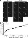
ipl1–321 mutants are defective in spindle disassembly. (A) Live microscopy was performed on wild-type (SBY130, left) and ipl1–321 mutant cells (SBY97, right) containing Tub1–GFP that were released from α-factor at 35°C. Eight z sections at 0.5-μm intervals were acquired every minute. Images of the spindle in a single cell are shown every 2 min after the initiation of anaphase (time 0'). An outline of the cell is shown at time 0'. Spindle disassembly is delayed in ipl1–321 cells, and the spindle orientation changes during the initial phases of anaphase. A hyperelongated spindle in an ipl1–321 mutant cell is shown in A*. See also videos 1 and 2 available at http://www.jcb.org/cgi/content/full/jcb.200209018/DC1. Bar, 10 μm. (B) The spindle length at each time point was measured, and the averages of 10 cells for each strain are graphed. Spindles disassemble in wild-type cells (○) 14 min after anaphase B initiation, whereas ipl1–321 mutant cells (▪) take 20 min.
Figure 4.
Figure 4.
Ipl1p's role in spindle disassembly is independent from its role in chromosome segregation. (A) pGAL-CDC20 (SBY952) and pGAL-CDC20 ipl1–321 (SBY943) cells containing Tub1–GFP were shifted to glucose to arrest cells in metaphase and then shifted to 37°C to inactivate Ipl1–321p. Cells were then released into galactose medium at 37°C to restore Cdc20 protein synthesis in the presence of α-factor to arrest cells in the following G1. The percentage of cells with a pole to pole distance ≥9 μm were monitored for the presence or absence of a spindle. Spindle disassembly occurred in 68% of pGAL-CDC20 cells compared with 36% of pGAL-CDC20 ipl1–321 mutant cells released from metaphase. (B) Wild-type (SBY130) and ipl1–321 (SBY97) cells containing Tub1–GFP were released from α-factor (T = 0) into the restrictive temperature (37°C). Time points were taken at 60, 70, and 80 min after release and monitored for the presence or absence of a spindle as in A. In wild-type cells, 78% of the spindles have depolymerized compared with only 40% of ip1l-321 mutant spindles. The bars represent the 95% confidence interval. (C) Cells from A were taken every 5 min, and Clb2p and Tub1p (loading control) protein levels were monitored by immunoblotting. Clb2p levels decline with similar kinetics in both strains, indicating that ipl1–321 mutant cells exit mitosis normally. (D) ipl1–321 mutants have hyperstable microtubules. cdc26Δ mcd1–1 (SBY965) and cdc26Δ mcd1–1 ipl1–321 (SBY2066) cells were arrested in G1 using α-factor then released into the cell cycle at the restrictive temperature (37°C). Cells were monitored for budding index, spindle formation, and spindle breakdown. 93% of cdc26Δ mcd1–1 mutant cells underwent spindle disassembly by 150 min compared with only 48% of cdc26Δ mcd1–1 ipl1–321 cells.
Figure 5.
Figure 5.
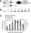
Ipl1p kinase activity increases before spindle disassembly. (A) Ip1lp was immunoprecipitated from a wild-type (SBY3) and a ipl1–321 mutant strain (SBY322) and then incubated with the histone-fold domain of the Cse4 kinetochore protein in a kinase reaction in vitro. The majority of Ipl1p present in the lysates before the immunoprecipitation (pre) was removed (post), and similar amounts of protein were used in the kinase assay (IP). The autoradiogram (right) shows that Cse4p is radiolabeled in the presence of wild-type Ipl1p but not Ipl1–321 mutant protein. (B) pGAL-CDC20 cells expressing Tub1–GFP (SBY952) were synchronized in metaphase by growth in glucose for 3 h. They were then released into galactose medium, and aliquots were taken every 10 min, and kinase assays were performed with the substrate Cse4p in vitro. The autoradiogram shows the phosphate incorporated into Cse4p in one experiment. (C) Microscopy was performed to determine the percent budding (▪) and the percent spindle disassembly (○). The total Ipl1p kinase activity is shown in gray bars (arbitrary units) for three experiments with the standard deviation indicated. Ipl1p kinase activity increases before spindle breakdown.
Figure 6.
Figure 6.
Ipl1p's substrates localize to the spindle midzone, and Ipl1p follows the plus ends of the depolymerizing spindle. (A) Microscopy was performed on strains containing endogenous Ndc10–GFP (SBY539), Sli15–GFP (SBY875), or Dam1–GFP (SBY1115). The fluorescence images show that all three Ipl1p substrates localize to the spindle midzone. The corresponding DIC pictures are shown (right). (B) Live image analysis was performed on cells expressing Ipl1–GFP and Tub1–CFP (SBY1036). Every 30 s, five z sections at 0.5-μm intervals were acquired while alternating between the two channels (FITC and CFP). The deconvolved video shows tubulin in red, Ipl1p in green, and the overlapping signal in yellow. Before spindle disassembly, Ipl1p localizes to the spindle midzone (0'). When the spindle starts breaking down (1'), the Ipl1p signal splits and then follows the plus ends of the depolymerizing spindle (2') until it reaches the spindle poles (3.5′). Video 3 showing Ipl1p localization is available at http://www.jcb.org/cgi/content/full/jcb.200209018/DC1. Bars, 10 μm.

Similar articles

Cited by

References

    1. Adams, R.R., S.P. Wheatleya, A.M. Gouldsworthy, S.E. Kandels-Lewis, M. Carmena, C. Smythe, D.L. Gerloff, and W.C. Earnshaw. 2000. INCENP binds the aurora-related kinase AIRK2 and is required to target it to chromosomes, the central spindle and cleavage furrow. Curr. Biol. 10:1075–1078. - PubMed
    1. Biggins, S., and A.W. Murray. 2001. The budding yeast protein kinase Ipl1/Aurora allows the absence of tension to activate the spindle checkpoint. Genes Dev. 15:3118–3129. - PMC - PubMed
    1. Biggins, S., F.F. Severin, N. Bhalla, I. Sassoon, A.A. Hyman, and A.W. Murray. 1999. The conserved protein kinase Ipl1 regulates microtubule binding to kinetochores in budding yeast. Genes Dev. 13:532–544. - PMC - PubMed
    1. Bischoff, J.R., L. Anderson, Y. Zhu, K. Mossie, L. Ng, B. Souza, B. Schryver, P. Flanagan, F. Clairvoyant, C. Ginther, et al. 1998. A homologue of Drosophila aurora kinase is oncogenic and amplified in human colorectal cancers. EMBO J. 17:3052–3065. - PMC - PubMed
    1. Chan, C.S., and D. Botstein. 1993. Isolation and characterization of chromosome-gain and increase-in-ploidy mutants in yeast. Genetics. 135:677–691. - PMC - PubMed

Publication types

MeSH terms