. 2003 Feb 3;160(3):355-64.
doi: 10.1083/jcb.200209022.

The role of the lissencephaly protein Pac1 during nuclear migration in budding yeast

Affiliations

The role of the lissencephaly protein Pac1 during nuclear migration in budding yeast

Wei-Lih Lee et al. J Cell Biol. .

Abstract

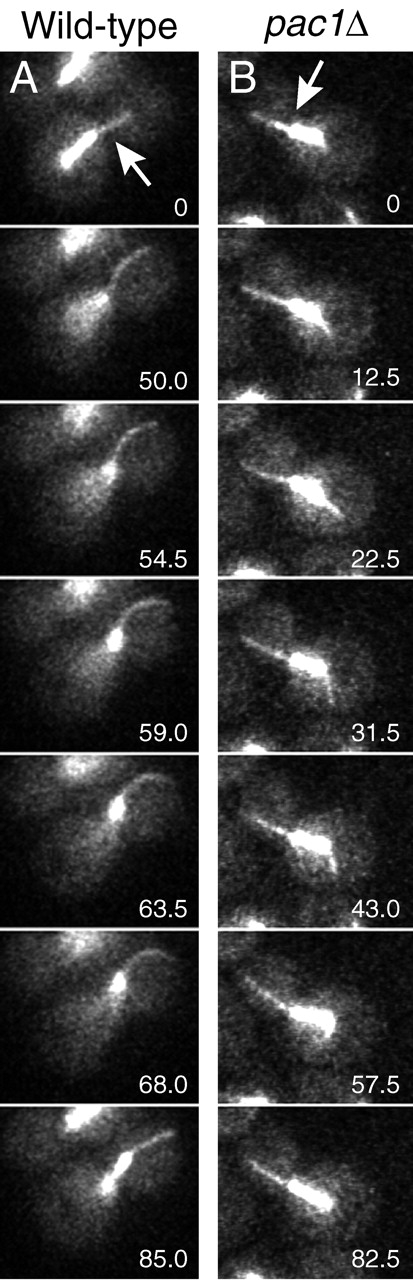
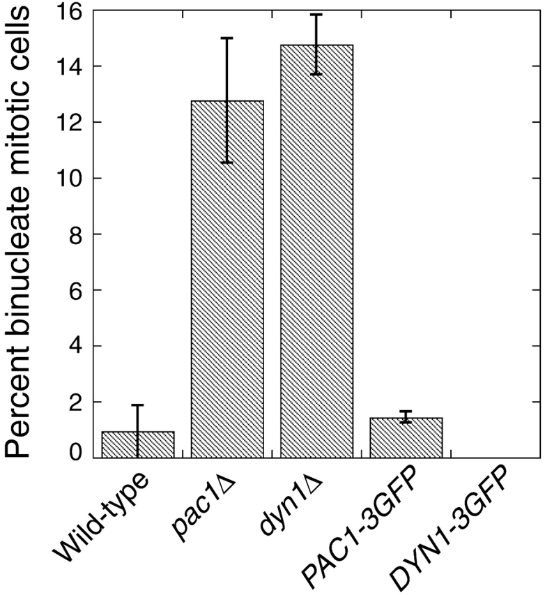
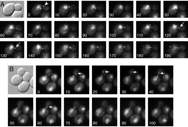
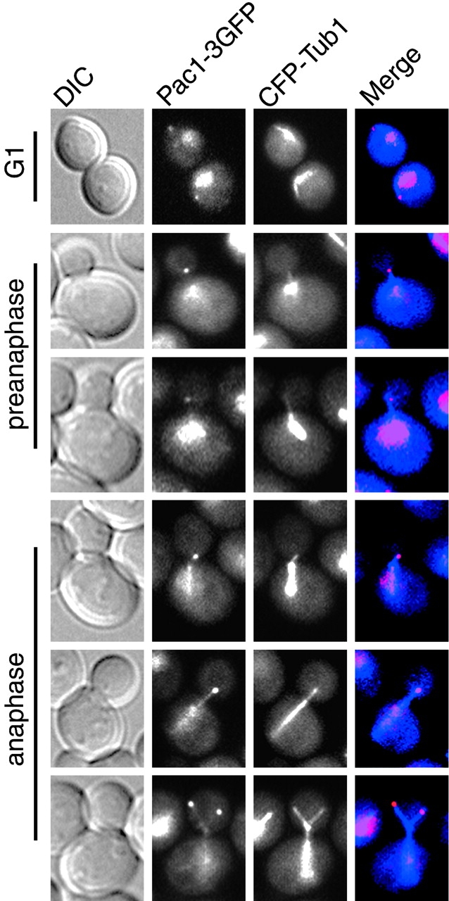
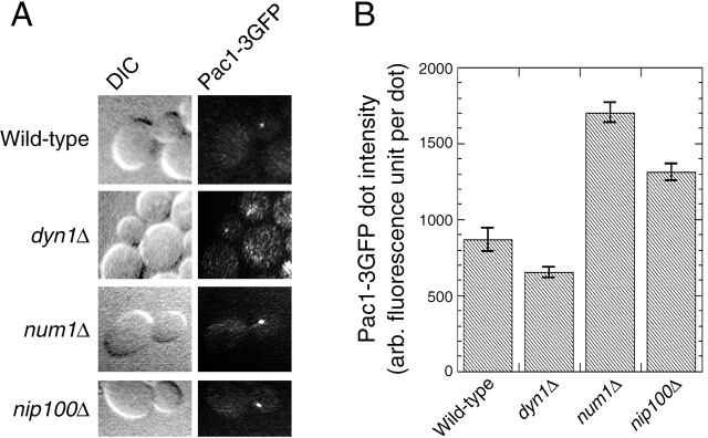
During mitosis in Saccharomyces cerevisiae, the mitotic spindle moves into the mother-bud neck via dynein-dependent sliding of cytoplasmic microtubules along the cortex of the bud. Here we show that Pac1, the yeast homologue of the human lissencephaly protein LIS1, plays a key role in this process. First, genetic interactions placed Pac1 in the dynein/dynactin pathway. Second, cells lacking Pac1 failed to display microtubule sliding in the bud, resulting in defective mitotic spindle movement and nuclear segregation. Third, Pac1 localized to the plus ends (distal tips) of cytoplasmic microtubules in the bud. This localization did not depend on the dynein heavy chain Dyn1. Moreover, the Pac1 fluorescence intensity at the microtubule end was enhanced in cells lacking dynactin or the cortical attachment molecule Num1. Fourth, dynein heavy chain Dyn1 also localized to the tips of cytoplasmic microtubules in wild-type cells. Dynein localization required Pac1 and, like Pac1, was enhanced in cells lacking the dynactin component Arp1 or the cortical attachment molecule Num1. Our results suggest that Pac1 targets dynein to microtubule tips, which is necessary for sliding of microtubules along the bud cortex. Dynein must remain inactive until microtubule ends interact with the bud cortex, at which time dynein and Pac1 appear to be offloaded from the microtubule to the cortex.


Figures

Figure 1.
Figure 1.
Microtubule and spindle behavior in wild-type and pac1 Δ strains. (A and B) Frames from movies of GFP-labeled microtubules during movement of the mitotic spindle into the mother–bud neck. Arrows indicate the position of the neck. The time elapsed in seconds is indicated. See Videos 1, 2, and 3 (available at http://www.jcb.org/cgi/content/full/jcb.200209022/DC1). Strains: wild-type GFP–TUB1, YJC2350; pac1Δ GFP–TUB1, YJC2501.
Figure 2.
Figure 2.
Function of PAC1–3GFP and DYN1–3GFP. Cells grown to mid-log phase at 12°C were fixed, stained for nuclei with DAPI, and imaged. The fraction of budded mitotic cells (Heil-Chapdelaine et al., 2000) with two nuclei in the mother is plotted. Error bars represent standard error (n > 750 cells counted for each strain). Strains: wild type, YJC2296; pac1Δ, YJC1629; dyn1Δ, YJC2007; PAC1–3GFP, YJC2770; DYN1–3GFP, YJC2772.
Figure 3.
Figure 3.
Localization of Pac1–3GFP. Differential interference contrast (DIC) and Pac1–3GFP wide-field fluorescence images of wild-type cells. (A) Pac1–3GFP is observed in the cytoplasm as dots (arrowhead), which move rapidly and sometimes form linear streaks (arrows). (B) Pac1–3GFP dots move toward and away from the bud (arrows). Pac1–3GFP is also observed in the nucleus, with a diffuse distribution. The time elapsed in seconds is indicated. Strain: PAC1–3GFP, YJC2770. See Videos 4 and 5 (available at http://www.jcb.org/cgi/content/full/jcb.200209022/DC1).
Figure 4.
Figure 4.
Pac1–3GFP colocalizes with the distal ends of cytoplasmic microtubules. DIC, Pac1–3GFP, and CFP–Tub1 wide-field fluorescence images of wild-type cells. The merged images on the right show Pac1–3GFP in red and CFP–Tub1 in blue. Cytoplasmic dots of Pac1–3GFP colocalize with the distal ends of microtubules at different stages of the cell cycle. Localization of Pac1–3GFP to microtubule ends was observed in the bud before the mitotic spindle moved into the neck (rows 4 and 5 from top) and after the mitotic spindle moved into the neck (row 6). Sometimes Pac1–3GFP was observed along cytoplasmic microtubules (rows 5 and 6). Strain: PAC1–3GFP CFP–TUB1, YJC2814.
Figure 5.
Figure 5.
Localization of Pac1–3GFP in living dyn1 Δ , num1 Δ , and nip100 Δ cells. (A) DIC and a frame from movies of Pac1–3GFP fluorescence in isogenic wild-type and mutant cells. The video camera and microscope settings were the same, allowing one to compare the intensity of fluorescence in the different strains. num1Δ and nip100Δ cells showed increased intensity of Pac1–3GFP dots in the bud. See Videos 6–9 (available at http://www.jcb.org/cgi/content/full/jcb.200209022/DC1). (B) Relative fluorescence intensity of motile Pac1–3GFP dots in wild-type and mutant cells. The average corrected fluorescence per dot is plotted; n = 25 dots for wild type, 75 dots for dyn1Δ, 170 dots for num1Δ, 171 dots for nip100Δ. Error bars represent standard error. Strains: PAC1–3GFP, YJC2770; PAC1–3GFP dyn1Δ, YJC2907; PAC1–3GFP num1Δ, YJC2905; PAC1–3GFP nip100Δ, YJC2904.
Figure 6.
Figure 6.
Localization of Dyn1–3GFP in living wild-type cells. (A) DIC and movie frames of Dyn1–3GFP fluorescence in a wild-type cell (YJC2772). Each fluorescence image is a two-dimensional projection of a 4-μm Z-stack of confocal images. The time elapsed in seconds is indicated. Dyn1–3GFP is observed as a dot that moves away from and toward the bud (arrows). Dyn1–3GFP sometimes appears as a linear streak (t = 28, 84, and 147 s). Dyn1–3GFP is also observed as stationary cortical dots, but only in the mother (see Videos 10 and 11, available at http://www.jcb.org/cgi/content/full/jcb.200209022/DC1). (B) Dyn1–3GFP colocalizes with the distal ends of cytoplasmic microtubules. DIC, Dyn1–3GFP, and CFP–Tub1 wide-field fluorescence images of wild-type cells (YJC2914) at G1 (top), preanaphase (middle), and anaphase (bottom). The merged images show cytoplasmic Dyn1–3GFP dots (red) at the distal ends of microtubules (blue).
Figure 7.
Figure 7.
Localization of Dyn1–3GFP in living arp1 Δ , num1 Δ , and pac1 Δ cells. (A) DIC and Dyn1–3GFP fluorescence images of isogenic wild-type and mutant cells. The video camera and microscope settings were the same for the different strains. arp1Δ and num1Δ cells showed increased fluorescence intensity for Dyn1–3GFP dots in the bud. pac1Δ cells showed the absence of cytoplasmic Dyn1–3GFP motile dots. See Videos 12–15 (available at http://www.jcb.org/cgi/content/full/jcb.200209022/DC1). (B) Relative fluorescence intensity of motile Dyn1–3GFP dots in wild-type, arp1Δ, and num1Δ cells. The average corrected fluorescence per dot is plotted; n = 72 dots for wild type, 91 dots for arp1Δ, 84 dots for num1Δ. Error bars represent standard error. Strains: DYN1–3GFP, YJC2772; DYN1–3GFP arp1Δ, YJC2908; DYN1–3GFP num1Δ, YJC2910; DYN1–3GFP pac1Δ, YJC2912.
Figure 8.
Figure 8.
Proposed mechanism for microtubule sliding along the bud cortex. (Step 1) Dynein Dyn1 (red) and Pac1 (yellow) associated with the distal plus end of a cytoplasmic microtubule probe the bud cortex for attachment sites, which contain Num1 (blue) and probably other components. (Step 2) Upon attachment to a cortical site, dynein and Pac1 are offloaded from the end of microtubule and anchored to the cortex. (Step 3) The motor activity of anchored dynein is activated, causing it to walk toward the SPB. The microtubule slides, and the spindle is pulled into the bud neck. See Video 16 (available at http://www.jcb.org/cgi/content/full/jcb.200209022/DC1) and text for further discussion.

Comment in

References

    1. Adames, N.R., and J.A. Cooper. 2000. Microtubule interactions with the cell cortex causing nuclear movements in Saccharomyces cerevisiae. J. Cell Biol. 149:863–874. - PMC - PubMed
    1. Amberg, D.C., J.E. Zahner, J.W. Mulholland, J.R. Pringle, and D. Botstein. 1997. Aip3p/Bud6p, a yeast actin-interacting protein that is involved in morphogenesis and the selection of bipolar budding sites. Mol. Biol. Cell. 8:729–753. - PMC - PubMed
    1. Baudin, A., O. Ozierkalogeropoulos, A. Denouel, F. Lacroute, and C. Cullin. 1993. A simple and efficient method for direct gene deletion in Saccharomyces cerevisiae. Nucleic Acids Res. 21:3329–3330. - PMC - PubMed
    1. Beach, D.L., J. Thibodeaux, P. Maddox, E. Yeh, and K. Bloom. 2000. The role of the proteins Kar9 and Myo2 in orienting the mitotic spindle of budding yeast. Curr. Biol. 10:1497–1506. - PubMed
    1. Carminati, J.L., and T. Stearns. 1997. Microtubules orient the mitotic spindle in yeast through dynein-dependent interactions with the cell cortex. J. Cell Biol. 138:629–641. - PMC - PubMed

Publication types

MeSH terms