. 2003 Feb 3;160(3):365-74.
doi: 10.1083/jcb.200209095.

Hierarchy of protein assembly at the vertex ring domain for yeast vacuole docking and fusion

Affiliations

Hierarchy of protein assembly at the vertex ring domain for yeast vacuole docking and fusion

Li Wang et al. J Cell Biol. .

Abstract

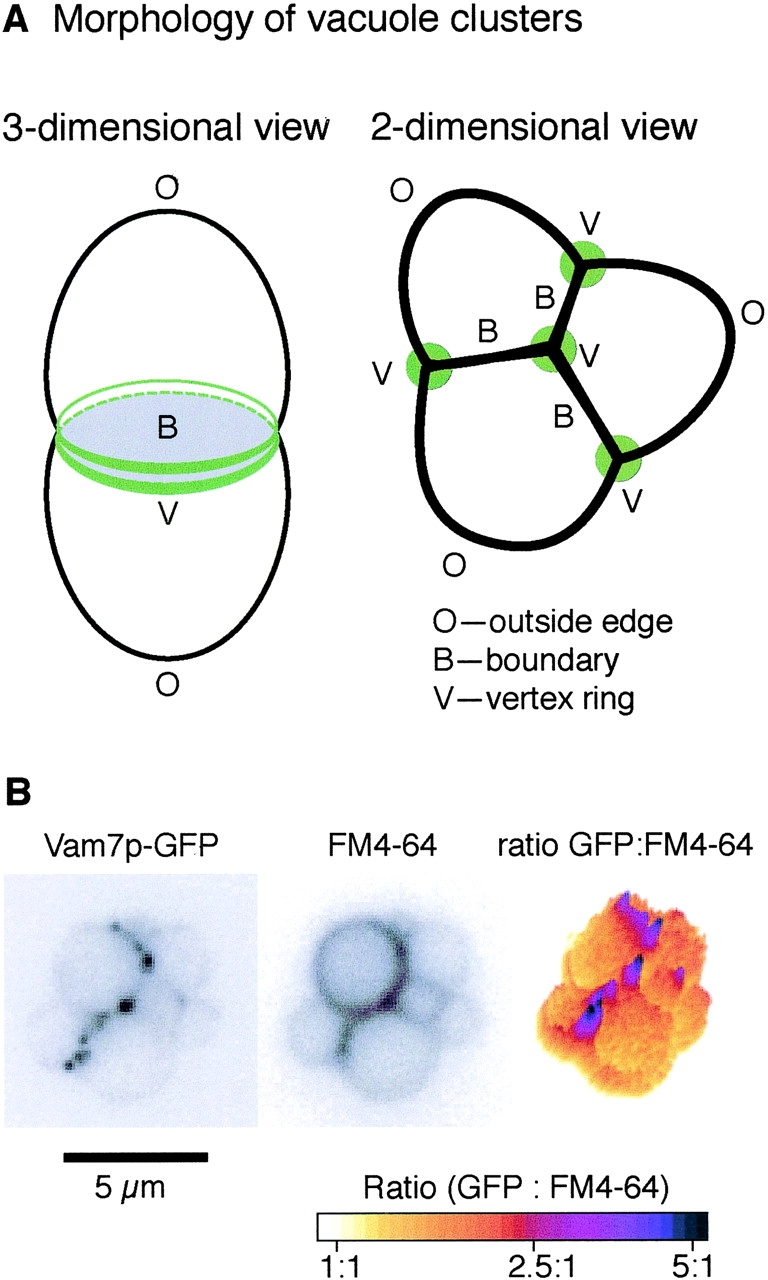
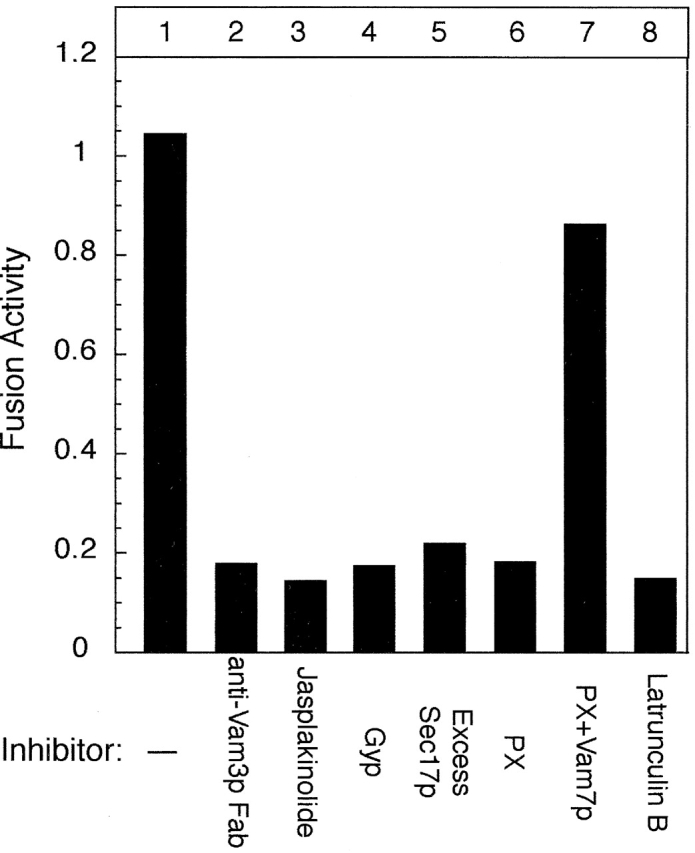
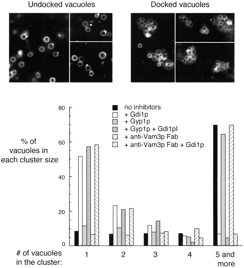
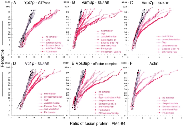
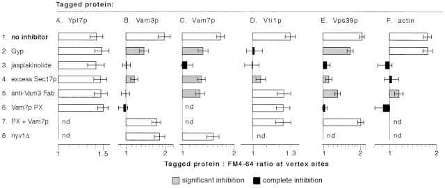
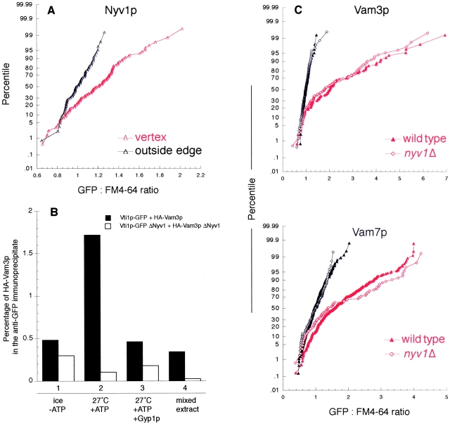
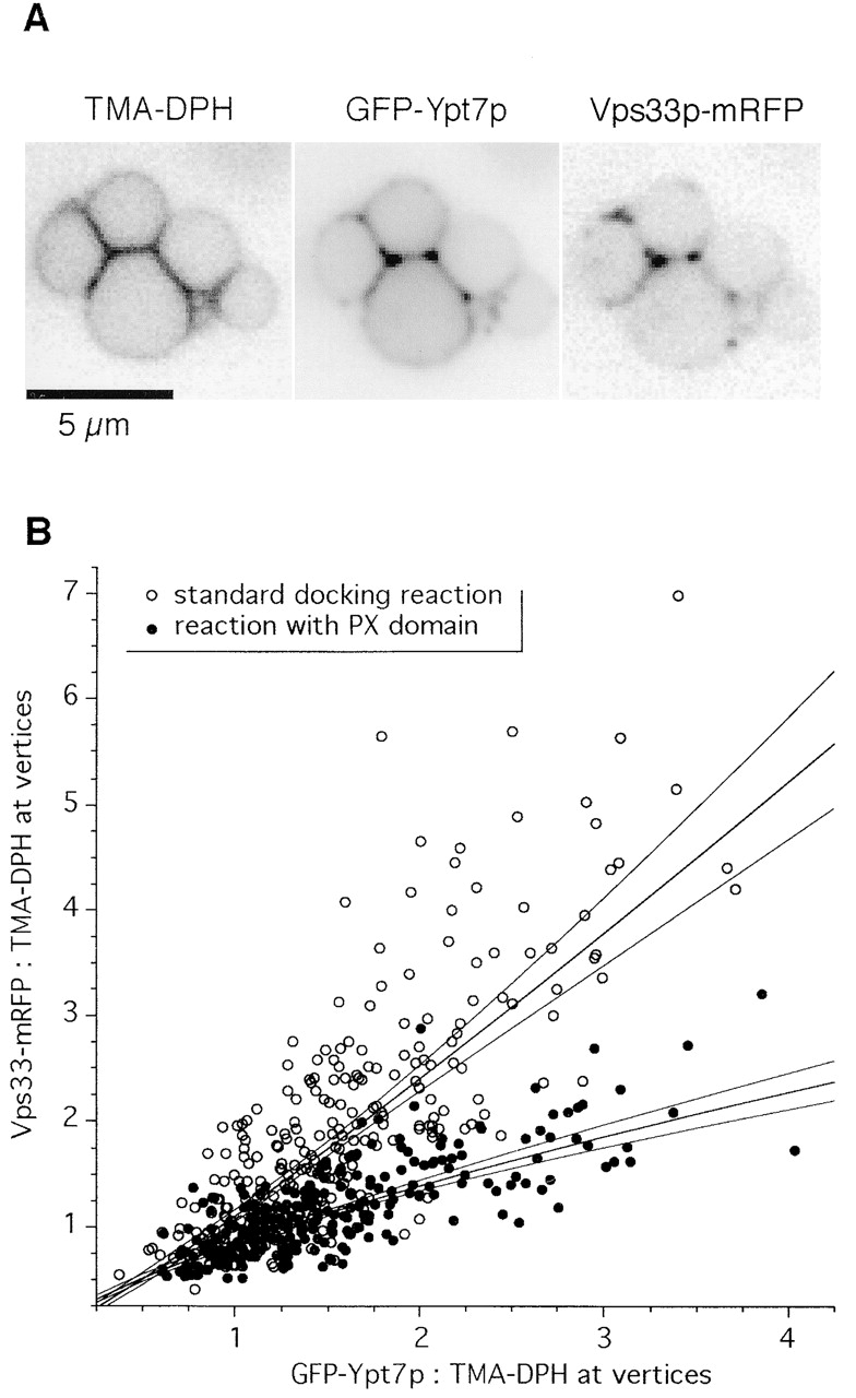
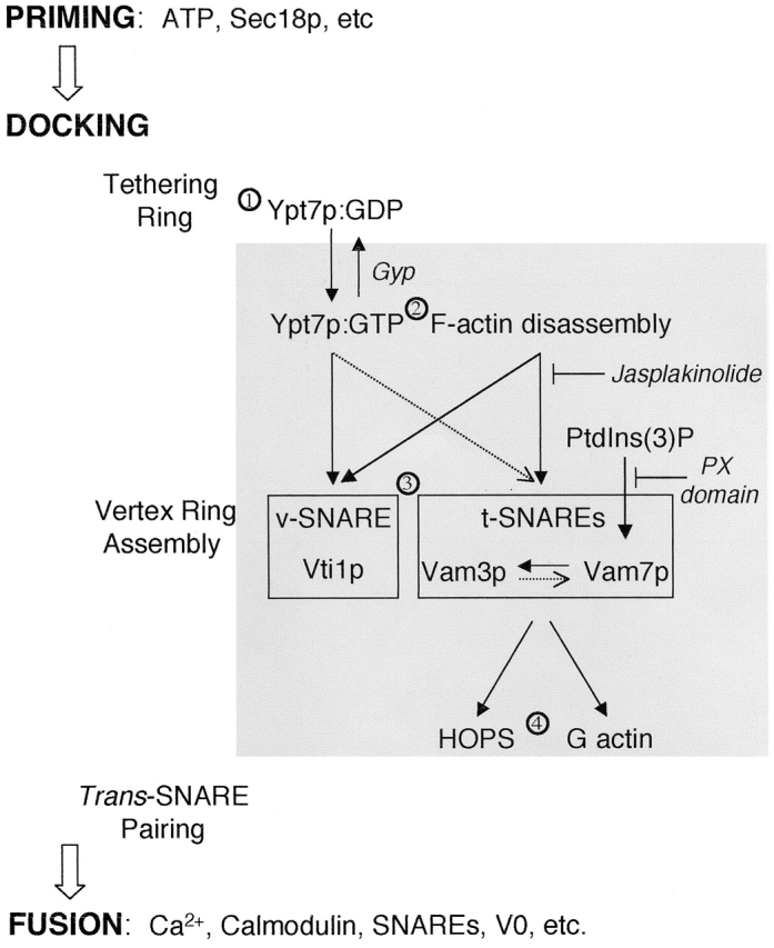
Vacuole tethering, docking, and fusion proteins assemble into a "vertex ring" around the apposed membranes of tethered vacuoles before catalyzing fusion. Inhibitors of the fusion reaction selectively interrupt protein assembly into the vertex ring, establishing a causal assembly hierarchy: (a) The Rab GTPase Ypt7p mediates vacuole tethering and forms the initial vertex ring, independent of t-SNAREs or actin; (b) F-actin disassembly and GTP-bound Ypt7p direct the localization of other fusion factors; (c) The t-SNAREs Vam3p and Vam7p regulate each other's vertex enrichment, but do not affect Ypt7p localization. The v-SNARE Vti1p is enriched at vertices by a distinct pathway that is independent of the t-SNAREs, whereas both t-SNAREs will localize to vertices when trans-pairing of SNAREs is blocked. Thus, trans-SNARE pairing is not required for SNARE vertex enrichment; and (d) The t-SNAREs regulate the vertex enrichment of both G-actin and the Ypt7p effector complex for homotypic fusion and vacuole protein sorting (HOPS). In accord with this hierarchy concept, the HOPS complex, at the end of the vertex assembly hierarchy, is most enriched at those vertices with abundant Ypt7p, which is at the start of the hierarchy. Our findings provide a unique view of the functional relationships between GTPases, SNAREs, and actin in membrane fusion.


Figures

Figure 1.
Figure 1.
In vitro vacuole tethering. (A) A schematic of tethered vacuoles. Membrane microdomains include outside edges (O), boundary membranes (B), and vertex membranes (V). (B) Vam7p is enriched at vertices. GFP-tagged Vam7p vacuoles were tethered in vitro (see Materials and methods) and labeled with FM4–64. Images were taken in GFP (left) and rhodamine (middle) channels; ratiometric images are on the right.
Figure 2.
Figure 2.
Catalytic assay of vacuole fusion. Vacuole fusion was assayed in vitro as described in Materials and methods. Fusion inhibitors, including 50 ng/μl excess Sec17p, 60 ng/μl anti-Vam3 Fab, 600 ng/μl recombinant Gyp1p, 500 μM jasplakinolide, 15 μM PX domain, 500 μM latrunculin B, and 5 μM full-length Vam7p were present where indicated.
Figure 3.
Figure 3.
Vacuole tethering. Inhibitors, such as 300 ng/μl Gdi1p, 600 ng/μl Gyp1p, and 60 ng/μl anti-Vam3p Fab were added as indicated to an in vitro docking assay. For each condition, images were acquired from random fields and an average of 500 vacuoles were scored for cluster size.
Figure 4.
Figure 4.
Protein enrichment at vertices of tethered vacuoles. Reaction inhibitors, including 50 ng/μl excess Sec17p, 60 ng/μl anti-Vam3 Fab, 600 ng/μl recombinant Gyp1p, 500 μM jasplakinolide, 15 μM PX domain, 500 μM latrunculin B, and 5 μM full-length Vam7p were added to docking assays (see Materials and methods) with GFP-tagged proteins as indicated. After incubation, vacuoles were observed by fluorescence microscopy and images were acquired in GFP and rhodamine channels. Vacuole clusters from random fields were analyzed and ratio values of GFP:FM4–64 were generated from outside edge membranes (black) and vertex membranes (red). An average of 200 normalized ratio values for each treatment were plotted against their percentile (cumulative distribution plot).
Figure 5.
Figure 5.
Statistical analysis of the effects of fusion inhibitors on protein enrichment at vertices. The bars show geometric means and 95% confidence intervals for the data sets that are shown as CD plots in Fig. 4. Shading indicates a treatment that causes statistically significant (P < 0.0006) partial or complete inhibition of vertex enrichment, compared with the corresponding “no inhibitor” condition (see Materials and methods).
Figure 6.
Figure 6.
Deletion of the v-SNARE Nyv1p has no effect on the localization of other SNAREs. (A) GFP-Nyv1p vacuoles were subject to in vitro docking assay and ratiometric fluorescence microscopy analysis, as described in Materials and methods. The GFP:FM4–64 ratio values were generated for outside edge membranes (red curves) and vertex membranes (black curves), and plotted against their percentile. (B) Trans-pairing of SNAREs. Trans-SNARE pairing was assayed directly using purified vacuoles from four strains: (1) VTI1-GFP, NYV1, pep4–3; (2) HA 3-VAM3, NYV1, pep4::HIS3; (3) VTI1-GFP, pep4::HIS3, nyv1::URA3; and (4) HA 3-VAM3, pep4::HIS3, nyv1::URA3. After a 60-min fusion reaction, membranes were dissolved in Triton X-100, and Vti1-GFP was immunoprecipitated by immobilized antibody to GFP (unpublished data). Trans-associated HA-Vam3p was detected by immunoblot and quantified by densitometry as a percentage of the total HA-Vam3p in the sample. Recombinant Gyp1–46p (0.4 μg/ml) was added where indicated (lane 3). To determine the level of background association, detergent extracts of reactions with ATP and single vacuole populations were prepared and mixed before immunoprecipitation (lane 4). (C) Vacuoles were isolated from nvy1Δ strains with GFP-tagged Vam3p or Vam7p and assayed for protein localization during docking as above (Fig. 4).
Figure 7.
Figure 7.
Ypt7p and Vps33p colocalization at vertex sites. GFP-Ypt7p and Vps33-mRFP were quantified at vertex sites under docking conditions in the absence (open circles) or presence (filled circles) of PX domain. Lines show linear fits to log-transformed intensity data and 99% confidence intervals for the best-fit lines. The upper fit is to the control samples (open circles) and the lower fit is to the PX domain-treated samples (filled circles). The data are pooled from three independent experiments; interexperimental variation did not significantly influence the results.
Figure 8.
Figure 8.
Working model of vertex ring assembly. See text for details. Solid black arrows indicate a strict hierarchical requirement for vertex enrichment. Dotted arrows indicate partial influences on vertex enrichment.

Similar articles

Cited by

References

    1. Albert, S., E. Will, and D. Gallwitz. 1999. Identification of the catalytic domains and their functionally critical arginine residues of two yeast GTPase-activating proteins specific for Ypt/Rab. EMBO J. 18:5216–5225. - PMC - PubMed
    1. Bennett, M.K., N. Calakos, and R.H. Scheller. 1992. Syntaxin: a synaptic protein implicated in docking of synaptic vesicles at presynaptic active zones. Science. 257:255–259. - PubMed
    1. Boeddinghaus, C., A.J. Merz, R. Laage, and C. Ungermann. 2002. A cycle of Vam7p release from and PtdIns 3-P-dependent rebinding to the yeast vacuole is required for homotypic vacuole fusion. J. Cell Biol. 157:79–89. - PMC - PubMed
    1. Bromley, S.K., W.R. Burack, K.G. Johnson, K. Somersalo, T.N. Sims, C. Sumen, M.M. Davis, A.S. Shaw, P.M. Allen, and M.L. Dustin. 2001. The immunological synapse. Annu. Rev. Immunol. 19:375–396. - PubMed
    1. Cao, X., N. Ballew, and C. Barlowe. 1998. Initial docking of ER-derived vesicles requires Uso1p and Ypt1p but is independent of SNARE proteins. EMBO J. 17:2156–2165. - PMC - PubMed

Publication types

MeSH terms