. 2003 Feb 1;17(3):394-409.
doi: 10.1101/gad.1044903.

Ectodermal Wnt3/beta-catenin signaling is required for the establishment and maintenance of the apical ectodermal ridge

Affiliations

Ectodermal Wnt3/beta-catenin signaling is required for the establishment and maintenance of the apical ectodermal ridge

Jeffery R Barrow et al. Genes Dev. .

Abstract

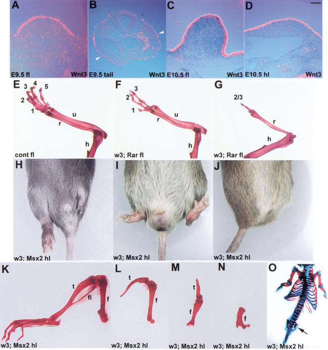
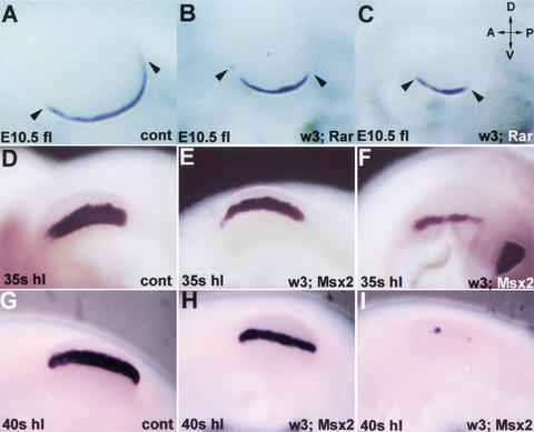
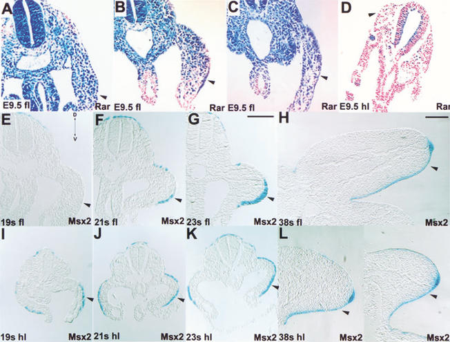
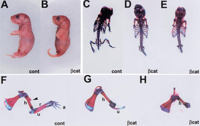
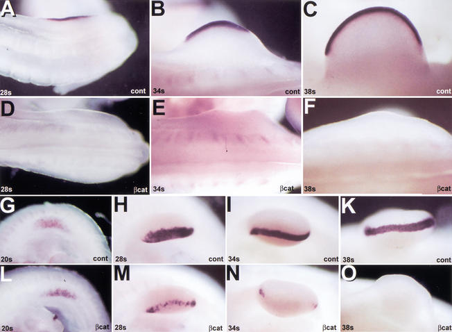
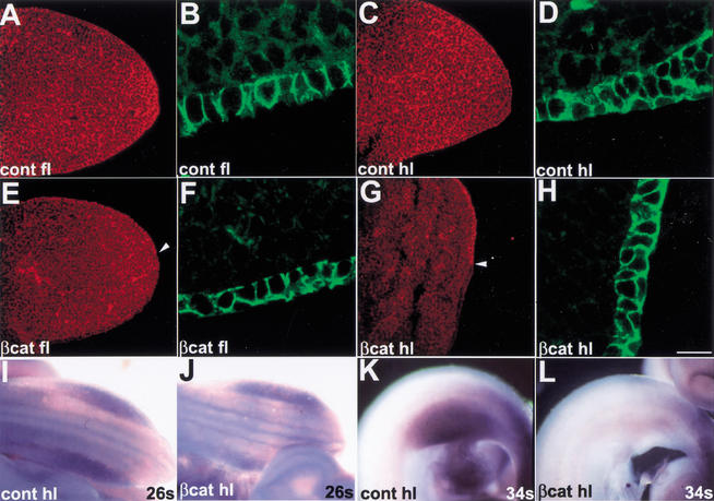
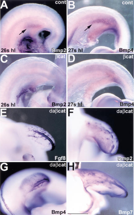
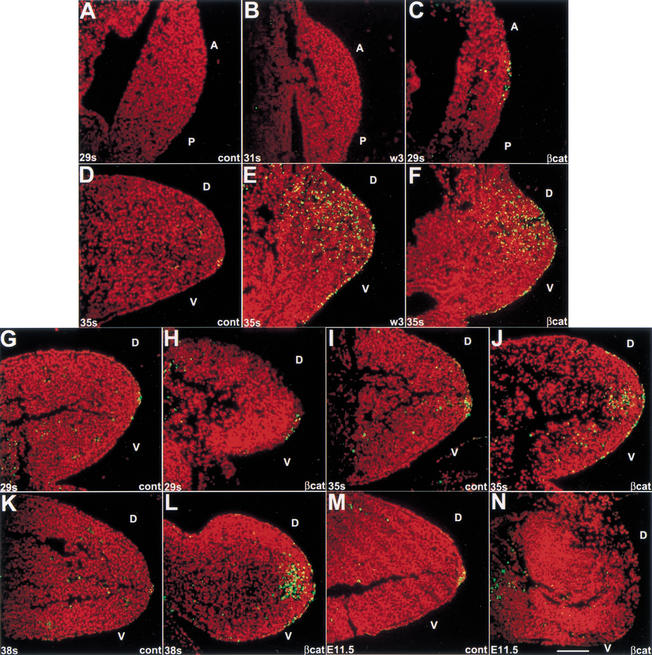
The formation of the apical ectodermal ridge (AER) is critical for the distal outgrowth and patterning of the vertebrate limb. Recent work in the chick has demonstrated that interplay between the Wnt and Fgf signaling pathways is essential in the limb mesenchyme and ectoderm in the establishment and perhaps the maintenance of the AER. In the mouse, whereas a role for Fgfs for AER establishment and function has been clearly demonstrated, the role of Wnt/beta-catenin signaling, although known to be important, is obscure. In this study, we demonstrate that Wnt3, which is expressed ubiquitously throughout the limb ectoderm, is essential for normal limb development and plays a critical role in the establishment of the AER. We also show that the conditional removal of beta-catenin in the ventral ectodermal cells is sufficient to elicit the mutant limb phenotype. In addition, removing beta-catenin after the induction of the ridge results in the disappearance of the AER, demonstrating the requirement for continued beta-catenin signaling for the maintenance of this structure. Finally, we demonstrate that Wnt/beta-catenin signaling lies upstream of the Bmp signaling pathway in establishment of the AER and regulation of the dorsoventral polarity of the limb.


Figures

Figure 1
Figure 1
Wnt3 expression and consequences of its removal in the limb ectoderm. (A) Wnt3 is expressed ubiquitously in the forelimb ectoderm at E9.5. (B) Transverse section through the tail at the level of the prospective hindlimbs at E9.5. Wnt3 is expressed at low levels throughout the ectoderm, including that overlying the prospective limb mesenchyme (arrowheads). (C,D) Wnt3 is expressed strongly throughout the ectoderm of both the fore- and hindlimbs at E10.5. (EO) Limb defects in Wnt3 conditional mutants. (EG) Wnt3n/c; RARCre mutants exhibit variable forelimb defects ranging from three digits (anterior digits 1–3) and normal zeugopod [radius (r) and ulna (u) in F] to one digit (digit 2 or 3) and also zeugopod defects (missing ulna in G). The humerus (h) in most instances was unaffected. (HO) The phenotype of Wnt3n/c; Msx2Cre conditional mutants varied dramatically, ranging from completely normal (H, right hindlimb; K) to entirely absent (arrow, O). cont, control animals; fl, forelimb; h, humerus; hl, hindlimb; numbers (i.e., 1–5) indicate digit number; r, radius; u, ulna; w3; Msx2, Wnt3n/c; Msx2Cre mutants; w3; Rar, Wnt3n/c; RARCre mutants; Bar: AD, 100 μm.
Figure 2
Figure 2
Fgf8 expression and AER formation are disrupted in Wnt3 conditional mutants. For AI, dorsal (D) is up, ventral is down, anterior (A) is left, and posterior (P) is right. (AC) Wnt3n/c; RARCre conditional mutants exhibit a reduction in the anterior/posterior length of the AER (arrowheads). Fgf8 expression in the RARCre mutant hindlimbs is normal. (DI) Wnt3n/c; Msx2Cre conditional mutants exhibit variability in the expression of Fgf8. E and H represent mutants where Fgf8 expression has been mildly affected, however, the dorsoventral girth of the AER is reduced to ∼50%. F and I represent variations of severely affected limbs such that Fgf8 expression is dramatically reduced. cont, control; fl, forelimb; hl, hindlimb; w3; Rar, Wnt3n/c; RARCre, w3; Msx, Wnt3n/c; Msx2Cre. Bar, 300 μm.
Figure 3
Figure 3
RARCre and Msx2Cre reporter activity in the limb ectoderm. For all panels ventral is down; accordingly, the arrowhead denotes the ventral ectoderm. (AD) ROSA26 reporter activity for RARCre transgenic fore- and hindlimbs. Sections through the anterior (A), central (B), and posterior (C) regions of the forelimb bud. Note the strong Cre activity throughout the ectoderm of the anterior (A) and posterior (C) sections, whereas the central section (B) only exhibits strong activity in the ventral ectoderm (arrowhead). (D) Transverse section through the hindlimb. (EL) ROSA26 reporter analysis of Msx2Cre transgenic fore- and hindlimbs. (E) At 19 somites (19s), Cre activity has not yet commenced in the forelimb ectoderm. (F) Cre is active weakly in the ventral ectoderm of 21s transgenic animals (arrowhead). (G,H) Cre activity is robust throughout the ventral ectoderm and AER. (I,L) Msx2Cre transgenic animals exhibit strong Cre activity throughout the ventral ectoderm of the hindlimbs (arrowheads). There is variable activity in the dorsal ectoderm. 19s, 19 somites; 20s, 20 somites; etc.; D, dorsal; fl, forelimb; hl, hindlimb; Msx2, Msx2Cre/R26R double transgenic animals; Rar, RARCre/R26R double transgenic embryos; V, ventral. Bars, 100 μm. Bar in G is for EG, and IK; bar in H is for H and L.
Figure 4
Figure 4
Limb defects in β-cateninn/c; Msx2Cre conditional mutant embryos. Control and mutant newborns (A,B) and newborn skeletons (CH). Note the complete absence of hindlimbs (B,D,E) and that the forelimbs are truncated at the end of the humerus (h; G,H). The proximal ulna (u) is present to variable extents (G,H), whereas the humerus is largely unaffected with the exception that the deltoid crest (arrowhead) is absent. a, autopod; βcat, β-cateninn/c; Msx2Cre; cont, control animals, r, radius; u, ulna.
Figure 5
Figure 5
Fgf8 expression is disrupted in the fore- and hindlimbs of β-cateninn/c; Msx2Cre conditional mutants. (AF) Dorsal views (anterior to the left) of control and mutant hindlimbs. Fgf8 is never expressed at any stage of development in the hindlimb ectoderm of β-cateninn/c; Msx2Cre mutants. (GO) Distal views (anterior to the left) of control and mutant forelimbs. Fgf8 expression initiates normally in the forelimb ventral ectoderm at 20 somites (L). It fades progressively at later stages (M,N) of development and is completely absent by 38 somites (O). cont, control; βcat, β-catenin; Msx2Cre; 20s, 28s, etc. refer to the age in somites of the embryo.
Figure 6
Figure 6
β-catenin removal in the limb ectoderm does not affect cell adhesion in the limb ectoderm nor induction of Fgf10 in the mesenchyme. (AH) Transverse sections (dorsal side up) through the fore- and hindlimbs of control and mutant embryos at 35 somites stained with β-catenin (rhodamine), and high-magnification views of ventral ectoderm from neighboring sections stained with E-cadherin (FITC) antibodies. In the control embryos (AD), both β-catenin and E-cadherin antibodies stain the membranes throughout the limb ectoderm. In the β-catenin; Msx2Cre mutants, however, β-catenin protein is restricted to the dorsal ectoderm (arrowheads, E,G). (F,H) Despite the absence of β-catenin in the ventral ectoderm, E-cadherin remains properly localized to the membranes of ventral ectodermal cells. Views of embryonic limb buds subjected to Fgf10 in situ hybridization. Note that early β-cateninn/c; Msx2Cre mutants exhibit normal Fgf10 expression (I,J), whereas at later stages Fgf10 is not maintained (K,L); arrows denote the position of the hindlimb. cont, control; βcat, β-catenin; Msx2Cre; Bar: A,C,E,G, 100 μm; B,D,F,H, 25 μm.
Figure 7
Figure 7
Ectodermal Wnt/β-catenin signaling lies upstream of Bmp signaling and dorsoventral patterning. Early expression of Bmp2 (A,C) and Bmp4 (B,D) in the ventral ectoderm (arrows, A,B) of the hindlimb is completely abolished in β-cateninn/c; Msx2Cre mutants (B,D). Activation of β-catenin in the chick limb ectoderm induces the expression of Fgf8 (E), Bmp2 (F), Bmp4 (G), and Bmp7 (H) in both the dorsal and ventral ectoderm. cont, control; βcat, β-catenin; Msx2Cre; daβcat, dominant active β-catenin; 26s, 27s, etc. refer to the age in somites of the embryos. Bar, 300 μm.
Figure 8
Figure 8
Lack of Wnt/β-catenin signaling in the limb ectoderm results in abnormal apoptosis in the ectoderm and mesenchyme of the limb. (AC) Apoptosis in the hindlimbs of control and mutants at 29–30 somites. There is no apoptosis in the limb mesenchyme of the control, Wnt3, or β-catenin mutants. There is, however, apoptosis in the ectoderm of the β-catenin mutants. (DF) At 35 somites (approximately a half-day after the induction of Fgf8), there is apoptosis in the AER of control embryos (D). In the Wnt3 and β-catenin conditional mutants, there is extensive apoptosis throughout the limb mesenchyme and adjacent ectoderm. The apoptosis in the mesenchyme is more significant dorsally. (GN) Apoptosis in the forelimbs of β-cateninn/c; Msx2Cre mutants. At 29 somites, there is apoptosis in the AER of controls (G) and in the AER-like thickened ectoderm of the β-catenin mutants (H). There is, however, no apoptosis in the limb mesenchyme. (J,L) At 35–38 somites, there is extensive apoptosis in the distal mesenchyme and associated ectoderm. (N) By E11.5 no ectopic apoptosis can be observed. 29s, 34s, etc., refer to the age in somites of the embryo; A, anterior; cont, control, βcat, β-catenin; Msx2Cre; D, dorsal; P, posterior; V, ventral; w3, Wnt3; Msx2Cre. Bar, 100 μm.

References

    1. Aberle H, Schwartz H, Kemler R. Cadherin–catenin complex: Protein interactions and their implications for cadherin function. J Cell Biochem. 1996;61:514–523. - PubMed
    1. Ahn K, Mishina Y, Hanks MC, Behringer RR, Crenshaw EB., III BMPR-IA signaling is required for the formation of the apical ectodermal ridge and dorsal-ventral patterning of the limb. Development. 2001;128:4449–4461. - PubMed
    1. Behrens J, von Kries JP, Kuhl M, Bruhn L, Wedlich D, Grosschedl R, Birchmeier W. Functional interaction of β-catenin with the transcription factor LEF-1. Nature. 1996;382:638–642. - PubMed
    1. Bell SM, Schreiner CM, Scott WJ. The loss of ventral ectoderm identity correlates with the inability to form an AER in the legless hindlimb bud. Mech Dev. 1998;74:41–50. - PubMed
    1. Boussadia O, Kutsch S, Hierholzer A, Delmas V, Kemler R. E-cadherin is a survival factor for the lactating mouse mammary gland. Mech Dev. 2002;115:53–62. - PubMed

Publication types