. 2003 Feb;111(3):347-55.
doi: 10.1172/JCI16306.

Endothelial lipase is a major determinant of HDL level

Affiliations

Endothelial lipase is a major determinant of HDL level

Tatsuro Ishida et al. J Clin Invest. 2003 Feb.

Abstract

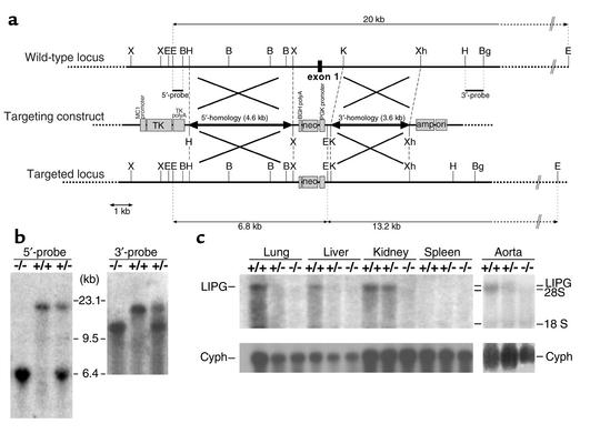
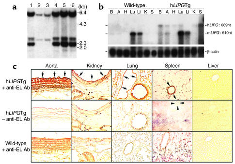
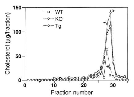
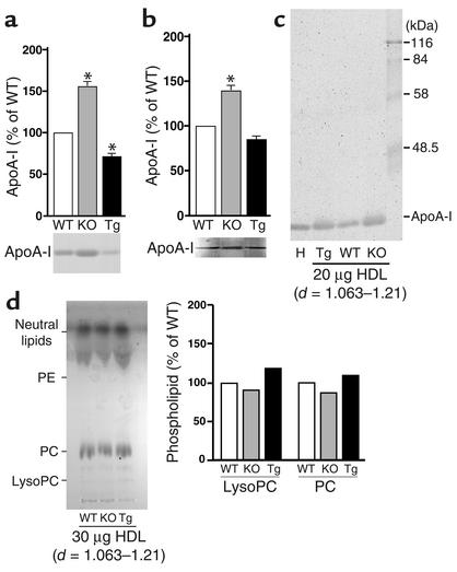
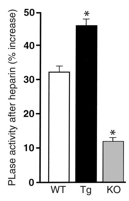
A new member of the lipase gene family, initially termed endothelial lipase (gene nomenclature, LIPG; protein, EL), is expressed in a variety of different tissues, suggesting a general role in lipid metabolism. To assess the hypothesis that EL plays a physiological role in lipoprotein metabolism in vivo, we have used gene targeting of the native murine locus and transgenic introduction of the human LIPG locus in mice to modulate the level of EL expression. Evaluation of these alleles in a C57Bl/6 background revealed an inverse relationship between HDL cholesterol level and EL expression. Fasting plasma HDL cholesterol was increased by 57% in LIPG(-/-) mice and 25% in LIPG(+/-) mice and was decreased by 19% in LIPG transgenic mice as compared with syngeneic controls. Detailed analysis of lipoprotein particle composition indicated that this increase was due primarily to an increased number of HDL particles. Phospholipase assays indicated that EL is a primary contributor to phospholipase activity in mouse. These data indicate that expression levels of this novel lipase have a significant effect on lipoprotein metabolism.


Figures

Figure 1
Figure 1
Targeting of the mouse endothelial lipase gene. (a) The wild-type locus of mouse LIPG (top), the targeting construct (middle), and the targeted locus (bottom). Exon 1 was replaced with the neomycin phosphotransferase gene (neo), and recombination was detected by Southern blot analysis using the probes indicated. X, XbaI; B, BamHI; K, KpnI; Bg, BglII; H, HindIII; E, EcoRI; Xh, XhoI. (b) Both probes gave a 20-kb wild-type band on Southern blotting, whereas the targeted recombinants showed a 6.8-kb band with the 5′probe and a 13.2-kb band with the 3′probe. Targeted stem cells were characterized by the presence of both DNA fragments. (c) Northern blot analysis showed LIPG expression in mouse tissues. A full-length mouse LIPG cDNA was used as a probe. Cyph, cyclophilin.
Figure 2
Figure 2
Generation of human endothelial lipase transgenic mice. (a) Genotyping of LIPG transgenic mice. Southern blot analysis was employed to detect the human LIPG transgene. Lanes 2 and 3 show wild-type mice; lanes 1, 4, and 6, heterozygous transgenic mice; and lane 5, homozygous transgenic mice. (b) RNase protection with hLIPGTg mouse RNAs. Human LIPG-specific signals were detected in the brain, aorta, heart, lung, kidney, and spleen. B, brain; A, aorta; H, heart; Lu, lung; Li, liver; K, kidney; S, spleen. (c) Immunohistochemical evaluation of human EL expression in transgenic mice. Tissues harvested from hLIPGTg mice were studied with an anti-human EL monoclonal antibody raised against a synthetic peptide. Human EL was primarily associated with the endothelium of the aorta and large vessels in the kidney, lung, and spleen as well as the microcirculation in the spleen and lung (arrows). Arrowheads in the spleen indicate the central artery, which does not stain when the primary antibody is omitted. No expression was detected in the liver. Original magnification: aorta, ×400; lung and liver, ×200; kidney and spleen, ×630.
Figure 3
Figure 3
Cholesterol levels in lipoprotein fractions obtained by FPLC. Determination of cholesterol concentration in serum fractions obtained by FPLC revealed a correlation between LIPG genotype and HDL cholesterol levels. HDL cholesterol levels for LIPG–/– mice were significantly greater than for wild-type mice in fractions 28 and 29 (P < 0.05). HDL cholesterol levels were lower in LIPG transgenic animals for fractions 29 and 30 (P < 0.05) than in wild-type mice. KO, LIPG–/–; Tg, hLIPGTg.*P < 0.05 vs. WT.
Figure 4
Figure 4
HDL lipoprotein particle analysis. (a) SDS-PAGE analysis of apoA-I in the HDL peak fractions isolated by FPLC. One hundred microliters of the HDL peak fraction from FPLC were evaluated by 15% SDS-PAGE. Gels were stained and quantitated by densitometry. (b) Western blot analysis of apoA-I in HDL peak fraction from FPLC. Images were quantitated by densitometry. (c) HDL isolated by density-gradient ultracentrifugation (d = 1.063–1.21) was evaluated by 12% SDS-PAGE, with 20 μg of protein loaded per lane. Lane 1 is human HDL standard, and lane 5 is a molecular weight marker. H, human HDL standard. (d) HDL isolated by density-gradient ultracentrifugation (d = 1.063–1.21) was evaluated by TLC. Equal amounts of HDL, as determined by protein quantitation, were loaded. KO, LIPG–/–; Tg, hLIPGTg. *P <0.05 compared to WT.
Figure 5
Figure 5
Phospholipase activity in genetic models. Phospholipase activity in different genetic lines was compared between pre- and postheparin injection samples. A significantly greater level of postheparin phospholipase activity was observed in the transgenic lines (*P < 0.05), and a significantly lower level of postheparin augmentation of phospholipase activity was found in the knockout animals (*P < 0.05) as compared to the wild-type controls. The preheparin levels of plasma phospholipase activity were as follows (mean ± SEM): wild-type animals, 519 ± 22.3 nmol FFA/ml/hr; knockout animals, 521 ± 5.4 nmol FFA/ml/hr; and transgenic animals, 510.5 ± 20.2 nmol FFA/ml/hr. PLase, phospholipase activity; Tg, hLIPGTg; KO, LIPG–/–.

Comment in

References

    1. Castelli WP, et al. HDL cholesterol and other lipids in coronary heart disease. The cooperative lipoprotein phenotyping study. Circulation. 1977;55:767–772. - PubMed
    1. Gordon T, Castelli WP, Hjortland MC, Kannel WB, Dawber TR. High density lipoprotein as a protective factor against coronary heart disease. The Framingham Study. Am. J. Med. 1977;62:707–714. - PubMed
    1. Heller DA, de Faire U, Pedersen NL, Dahlen G, McClearn GE. Genetic and environmental influences on serum lipid levels in twins. N. Engl. J. Med. 1993;328:1150–1156. - PubMed
    1. Hasstedt SJ, et al. The inheritance of high density lipoprotein cholesterol and apolipoproteins A-I and A-II. Atherosclerosis. 1984;51:21–29. - PubMed
    1. Rader DJ, Ikewaki K. Unravelling high density lipoprotein-apolipoprotein metabolism in human mutants and animal models. Curr. Opin. Lipidol. 1996;7:117–123. - PubMed

Publication types

MeSH terms