

Drosophila DEG/ENaC pickpocket genes are expressed in the tracheal system, where they may be involved in liquid clearance
- PMID: 12571352
- PMCID: PMC149970
- DOI: 10.1073/pnas.252785099
Drosophila DEG/ENaC pickpocket genes are expressed in the tracheal system, where they may be involved in liquid clearance
Abstract
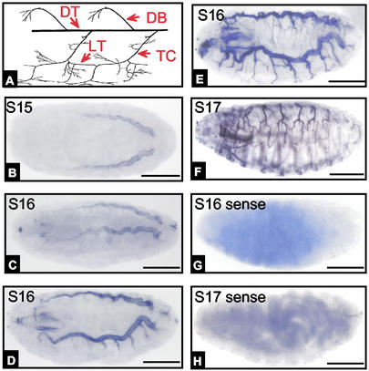
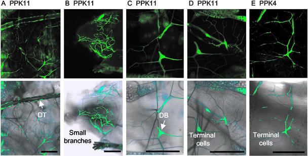
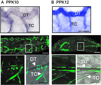
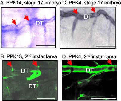
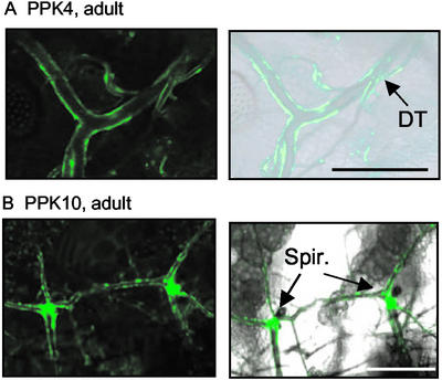
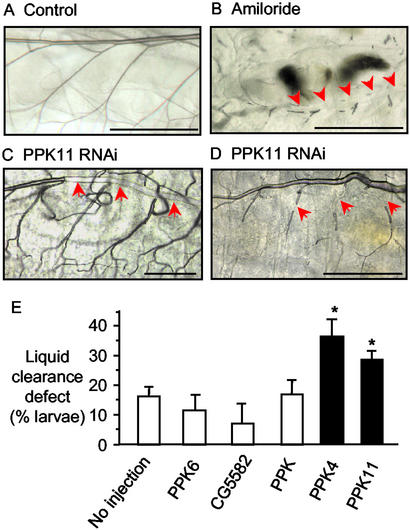
The Drosophila tracheal system and mammalian airways are branching networks of tubular epithelia that deliver oxygen to the organism. In mammals, the epithelial Na(+) channel (ENaC) helps clear liquid from airways at the time of birth and removes liquid from the airspaces in adults. We tested the hypothesis that related Drosophila degenerin (DEG)/ENaC family members might play a similar role in the fly. Among 16 Drosophila DEG/ENaC genes, called pickpocket (PPK) genes, we found 9 expressed in the tracheal system. By in situ hybridization, expression appeared in late-stage embryos after tracheal tube formation, with individual PPK genes showing distinct temporal and spatial expression patterns as development progressed. Promoters for several PPK genes drove reporter gene expression in the larval and adult tracheal systems. Adding the DEG/ENaC channel blocker amiloride to the medium inhibited liquid clearance from the trachea of first instar larvae. Moreover, when RNA interference was used to silence PPK4 and PPK11, larvae failed to clear tracheal liquid. These data suggest substantial molecular diversity of DEG/ENaC channel expression in the Drosophila tracheal system where the PPK proteins likely play a role in Na(+) absorption. Extensive similarities between Drosophila and mammalian airways offer opportunities for genetic studies that may decipher further the structure and function of DEG/ENaC proteins and development of the airways.
Figures
References
-
- Manning G, Krasnow M A. In: The Development of Drosophila melanogaster. Bate M, editor. Vol. 1. New York: Cold Spring Harbor Lab. Press; 1993. pp. 609–685.
-
- Metzger R J, Krasnow M A. Science. 1999;284:1635–1639. - PubMed
-
- Garty H, Palmer L G. Physiol Rev. 1997;77:359–396. - PubMed
-
- Alvarez de la Rosa D, Canessa C M, Fyfe G K, Zhang P. Annu Rev Physiol. 2000;62:573–594. - PubMed
-
- Hummler E, Barker P, Gatzy J T, Beermann F, Verdumo C, Schmidt A, Boucher R C J, Rossier B C. Nat Genet. 1996;12:325–328. - PubMed
Publication types
MeSH terms
Substances
Associated data
- Actions
- Actions
- Actions
- Actions
- Actions
- Actions
- Actions
- Actions
- Actions
- Actions
- Actions
- Actions
- Actions
- Actions
- Actions
- Actions
- Actions
- Actions
Grants and funding
LinkOut - more resources
Full Text Sources
Molecular Biology Databases
