. 2003 Feb 18;100(4):1879-84.
doi: 10.1073/pnas.0237137100. Epub 2003 Feb 5.

STAT3 deletion during hematopoiesis causes Crohn's disease-like pathogenesis and lethality: a critical role of STAT3 in innate immunity

Affiliations

STAT3 deletion during hematopoiesis causes Crohn's disease-like pathogenesis and lethality: a critical role of STAT3 in innate immunity

Thomas Welte et al. Proc Natl Acad Sci U S A. .

Abstract

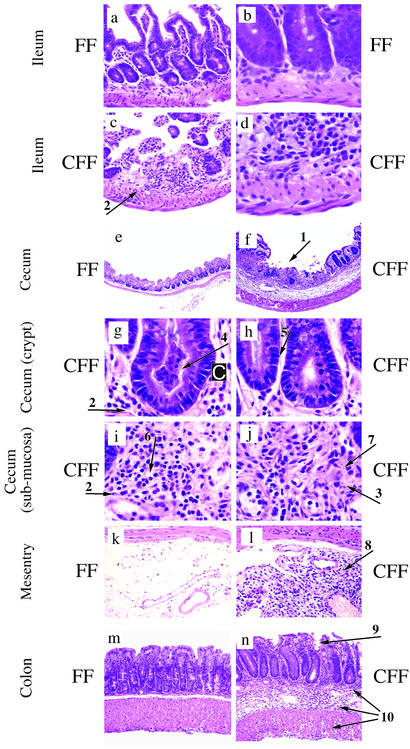
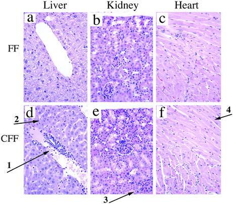
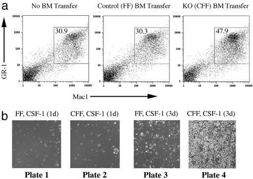
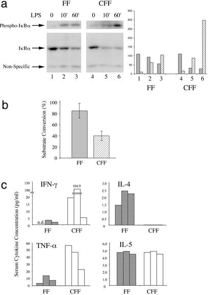
Signal transducer and activator of transcription 3 (STAT3) is a key transcriptional mediator for many cytokines and is essential for normal embryonic development. We have generated a unique strain of mice with tissue-specific disruption of STAT3 in bone marrow cells during hematopoiesis. This specific STAT3 deletion causes death of these mice within 4-6 weeks after birth with Crohn's disease-like pathogenesis in both the small and large intestine, including segmental inflammatory cell infiltration, ulceration, bowel wall thickening, and granuloma formation. Deletion of STAT3 causes significantly increased cell autonomous proliferation of cells of the myeloid lineage, both in vivo and in vitro. Most importantly, Stat3 deletion during hematopoiesis causes overly pseudoactivated innate immune responses. Although inflammatory cytokines, including tumor necrosis factor alpha and IFN-gamma, are overly produced in these mice, the NAPDH oxidase activity, which is involved in antimicrobial and innate immune responses, is inhibited. The signaling responses to lipopolysaccharide are changed in the absence of STAT3, leading to enhanced NF-kappa B activation. Our results suggest a model in which STAT3 has critical roles in the development and regulation of innate immunity, and deletion of STAT3 during hematopoiesis results in abnormalities in myeloid cells and causes Crohn's disease-like pathogenesis.


Figures

Figure 1
Figure 1
STAT3 deficiency during hematopoiesis results in Crohn's disease-like pathology. (aj) Ulcers (arrow 1), transmural infiltration (arrow 2), and granuloma-like structures (arrow 3) in the ileocecal area of STAT3-CFF mice in c, d, and fj. For comparison, the normal phenotype is depicted in a, b, and e. (c and d) Ileum of STAT3-CFF mouse. (e) Cecum of a control littermate and (f) of a STAT3-CFF mouse. (Magnifications: ×40, ad; ×10, e and f.) (gj) Higher-magnification views (×100, hematoxylin and eosin). (g) Crypt abscesses with necrotic neutrophils and monocytes (arrow 4), (h) high frequency of mitosis in the epithelium (arrow 5), (i) a marked infiltration of neutrophils, macrophages, and eosinophils (arrow 6) and few lymphocytes, edema of the lamina propria, (j) epithelioid cells with eosinophilic cytoplasm (arrow 7) and other inflammatory cells in the lamina propria can be identified. (k) Intestinal mesentry of control and (l) STAT3-deleted mice. Infiltration of neutrophils, macrophages, eosinophils (arrow 8 in l), and other cell types in the mesenteric fat tissue of STAT3-CFF mouse (×40, hematoxylin and eosin). Six sex-matched pairs of control (STAT3-FF) and STAT3-CFF littermates between 4 and 6 weeks of age were analyzed with similar results. IBD also spreads to the colon (n). Note bowel wall thickening with mucosal erosion (arrow 9) and transmural inflammation (arrow 10) in STAT3-CFF mice (×40, hematoxylin and eosin).
Figure 2
Figure 2
The inflammatory phenotype is not restricted to the gut but also occurs in liver. (ac) Tissue sections of liver, kidney, and heart of control littermates. (Magnifications: ×40.) (df) STAT3-CFF littermates. (d) The liver of STAT3-CFF mice was infiltrated around the portal vein by neutrophils, monocytes, and other cell types (arrow 1); hepatocytes were of normal appearance (arrow 2) (×40, hematoxylin and eosin). (e) Kidney had normal glomeruli (arrow 3) and tubular structures without inflammatory cell infiltration (×40, hematoxylin and eosin). (f) Heart showed normal myocardial fibers (arrow 4) without inflammatory cell infiltration (×40, hematoxylin and eosin). Five age- and sex-matched pairs were analyzed. Pathologic alteration of the liver was observed in all STAT3-CFF animals examined.
Figure 3
Figure 3
In the absence of STAT3, hematopoietic development is skewed toward the myeloid lineage at an early stage. (a) The presence of the myeloid lineage (GR-1+Mac1+) and erythroid lineage (TER119+) in the bone marrow of sex-matched littermates was analyzed by FACS staining. The genotype of the control is STAT3-FF. Heterozygous is STAT3-CF+ and knockout (KO) is STAT3-CFF. A result with typical ratios of GR-1+Mac1+ and TER119+ cells is shown. Three repeats with heterozygous animals and at least 15 repeats with control and STAT3-CFF mice gave similar ratios between the individual cell lineages. (b) Bone marrow (BM) was cultured for 7 days in methyl-cellulose in the presence of cytokines (IL-3, IL-6, stem cell factor, and insulin). The numbers of colonies formed were counted. Results obtained with four sex-matched pairs of control (STAT3-FF) and STAT3-CFF littermates are shown. The difference in the colony numbers of control and STAT3-CFF mice was not statistically significant. (c) FACS analyses with antibodies against c-Kit and different lineage markers (Mac1, B220, CD4, CD8, and TER119). The percentage of c-Kit+ lineage marker cells in the bone marrow of STAT3-CFF mice was evaluated and compared with sex-matched littermates. The results of five independent experiments are shown.
Figure 4
Figure 4
STAT3 regulates the generation of the myeloid lineage in a cell autonomous fashion in vivo and in vitro. (a) Bone marrow (BM) was transferred from a donor with control genotype (Center, STAT3-FF) or from a STAT3-CFF donor (Right) into lethally irradiated recipients. The presence of donor-derived bone marrow was determined after 8 weeks. Bone marrow was analyzed for the presence of GR-1+Mac1+ cells and compared with bone marrow of an age-matched untreated animal (Left). Experiments with three controls and three STAT3-CFF animals showed an increased percentage of GR-1+Mac1+ cells in the absence of STAT3. (b) Bone marrow from control (plates 1 and 3, STAT3-FF) and STAT3-deleted (plates 2 and 4, STAT3-CFF) littermates was cultured for 1 and 3 days in the presence of CSF-1 (30 ng/ml). Light microscopic pictures were taken showing a dramatic increase in the generation of attached cells in cultures of STAT3-deficient bone marrow. FACS with Mac1 antibody confirmed that these cells were macrophages. Three repeats showed similar results. (Magnification: ×2.)
Figure 5
Figure 5
STAT3 regulates crucial functional responses of the innate immune system. (a) IκB protein and phosphorylation levels were measured by Western blotting with specific antibodies to IκB and phospho-IκB before and after 10-min and 1-h LPS (10 ng/ml) stimulation of bone marrow-derived myeloid cells cultured in granulocyte/macrophage-CSF as indicated (FF, control mice; CFF, STAT3-CFF mice). (b) NADPH oxidase activity was measured in neutrophils isolated from whole blood samples of 4- to 5-week-old mice. Five controls (shaded bar) were compared with five sex-matched littermates (hatched bars). The difference between control and STAT3 knockout mice was statistically significant (five pairs analyzed). P value of Student's t test was P < 0.004. (c) Sera from 4- to 5-week-old mice were collected. IFN-γ, IL-4, tumor necrosis factor α (TNF-α), and IL-5 levels were measured. The results with three matched pairs of control (shaded bars) and STAT3-CFF littermates (open bars) are shown.

References

    1. Medzhitov R, Janeway C A., Jr Cell. 1997;91:295–298. - PubMed
    1. Strober W, Fuss I J, Blumberg R S. Annu Rev Immunol. 2002;20:495–549. - PubMed
    1. Duchmann R, Kaiser I, Hermann E, Mayet W, Ewe K, Meyer zum Buschenfelde K H. Clin Exp Immunol. 1995;102:448–455. - PMC - PubMed
    1. Hugot J P, Zouali H, Lesage S, Thomas G. Int J Colorectal Dis. 1999;14:2–9. - PubMed
    1. Laroux F S, Pavlick K P, Wolf R E, Grisham M B. News Physiol Sci. 2001;16:272–277. - PubMed

Publication types