

The crystallographic model of rhodopsin and its use in studies of other G protein-coupled receptors
- PMID: 12574068
- PMCID: PMC1351250
- DOI: 10.1146/annurev.biophys.32.110601.142520
The crystallographic model of rhodopsin and its use in studies of other G protein-coupled receptors
Abstract
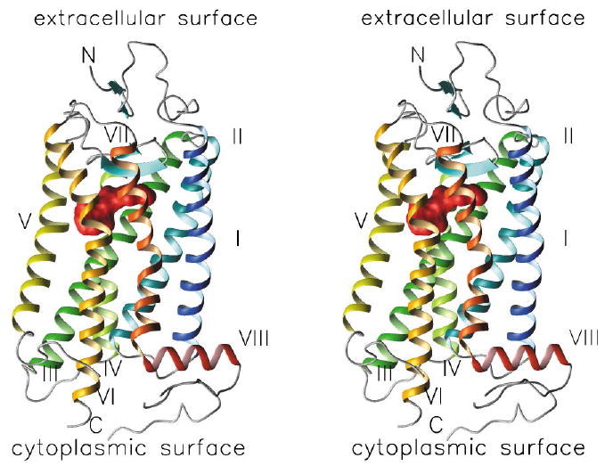
G protein-coupled receptors (GPCRs) are integral membrane proteins that respond to environmental signals and initiate signal transduction pathways activating cellular processes. Rhodopsin is a GPCR found in rod cells in retina where it functions as a photopigment. Its molecular structure is known from cryo-electron microscopic and X-ray crystallographic studies, and this has reshaped many structure/function questions important in vision science. In addition, this first GPCR structure has provided a structural template for studies of other GPCRs, including many known drug targets. After presenting an overview of the major structural elements of rhodopsin, recent literature covering the use of the rhodopsin structure in analyzing other GPCRs will be summarized. Use of the rhodopsin structural model to understand the structure and function of other GPCRs provides strong evidence validating the structural model.
Figures


References
-
- Albert AD, Watts A, Spooner P, Groebner G, Young J, Yeagle PL. A distance measurement between specific sites on the cytoplasmic surface of bovine rhodopsin in rod outer segment disk membranes. Biochim Biophys Acta. 1997;1328:74–82. - PubMed
-
- Altenbach C, Cai KW, Klein-Seetharaman J, Khorana HG, Hubbell WL. Structure and function in rhodopsin: mapping light-dependent changes in distance between residue 65 in helix TM1 and residues in the sequence 306–319 at the cytoplasmic end of helix TM7 and in helix H8. Biochemistry. 2001;40:15483–92. - PubMed
-
- Altenbach C, Klein-Seetharaman J, Cai KW, Khorana HG, Hubbell WL. Structure and function in rhodopsin: mapping light-dependent changes in distance between residue 316 in helix 8 and residues in the sequence 60–75, covering the cytoplasmic end of helices TM1 and TM2 and their connection loop CL1. Biochemistry. 2001;40:15493–500. - PubMed
-
- Ascoli M, Fanelli F, Segaloff DL. The lutropin/choriogonadotropin receptor, a 2002 perspective. Endocr Rev. 2002;23:141–74. - PubMed
-
- Baldwin JM, Schertler GFX, Unger VM. An alpha-carbon template for the transmembrane helices in the rhodopsin family of G-protein-coupled receptors. J Mol Biol. 1997;272:144–64. - PubMed
Publication types
MeSH terms
Substances
Grants and funding
LinkOut - more resources
Full Text Sources