. 2003 Feb 17;22(4):826-32.
doi: 10.1093/emboj/cdg095.

Galpha(s) is palmitoylated at the N-terminal glycine

Affiliations

Galpha(s) is palmitoylated at the N-terminal glycine

Christiane Kleuss et al. EMBO J. .

Abstract

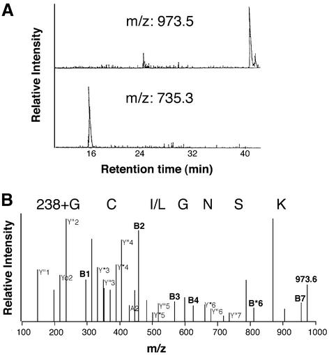
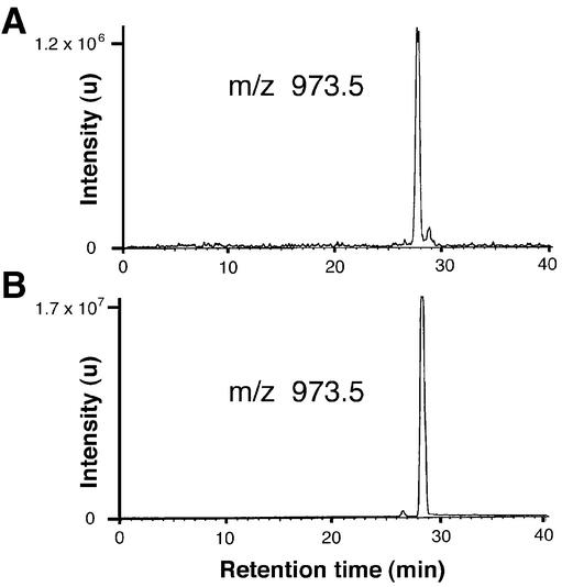
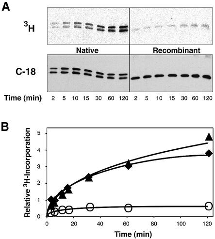
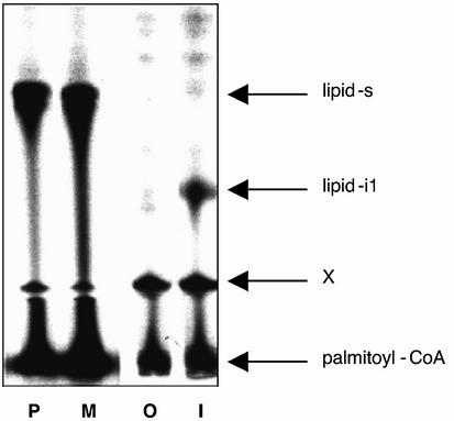
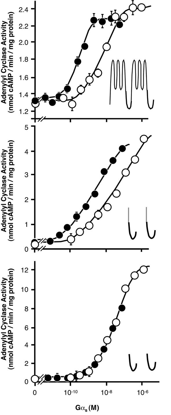
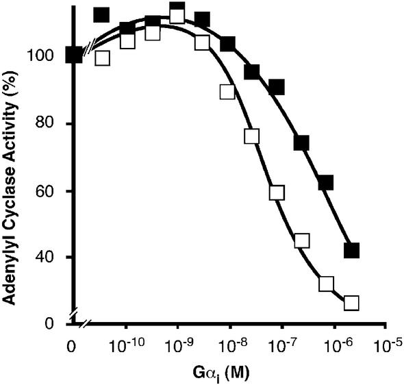
Covalent lipid attachments are essential co- and post-translational modifications for signalling proteins. Galpha(s), the alpha-subunit of the heterotrimeric G protein that activates adenylyl cyclase, is known to be palmitoylated at the third N-terminal amino acid, a cysteine. Palmitoylation is involved in anchoring Galpha(s) to the membrane by increasing its intrinsic hydrophobicity. We identified by mass spectrometry a second, functionally even more important, covalent modification. It consists of another palmitoyl residue attached to the preceding glycine (Gly(2)). Palmitoylation at this position has profound consequences for levels of signal transduction. It sensitizes the cell up to 200-fold for adenylyl cyclase-stimulating agents. The inhibitory inputs mediated by Galpha(i) are downregulated to <10%. Thereby, Gly(2)-palmitoylation of Galpha(s) relieves cellular stimulation at the level of adenylyl cyclase whereas it renders the inhibitory modulation via Galpha(i) more difficult.


Figures

None
Fig. 1. Mass spectrometric analysis of Gαs. Native or recombinant Gαs was in-gel digested and the resulting peptides analysed by MALDI-MS. Mass peaks with m/z 973.5 and 735.3 corresponded to the palmitoylated and non-palmitoylated N-terminal sequence G2CLGNSK of Gαs, respectively. (A) LC chromatogram recorded for the native protein at m/z 973.5 and the recombinant protein at m/z 735.3. (B) MS/MS spectrum of m/z = 973.58 of native Gαs. Relevant ions were labelled according to the accepted nomenclature and listed in Table II.
None
Fig. 2. LC/MS analysis. Mass chromatogram (m/z 973.5) of the peptide mixture generated by in-gel digestion of native Gαs with Lys-C (A) and of the peptide palm-GCLGNSK prepared by solid-phase peptide synthesis (B). Samples were separated by HPLC and detected on-line by electrospray mass spectrometry. Indicated are signal intensities in arbitrary units (u).
None
Fig. 3. Autopalmitoylation of Gαs at Cys3. Gαs (2.5 µM) purified from recombinant bacteria (open circle) or native source (filled triangle for Gαs-short, filled diamond for Gαs-long) were incubated with 25 µM [9,10–3H]palmitoyl–coenzyme A (20 000 c.p.m./pmol) for the indicated times at 20°C. (A) Shown are 3H-images of reaction products after electrophoretic separation and transfer onto nitrocellulose membranes (upper panel), and immunostains with Gαs-specific antiserum C-18 (lower panel). (B) Relative 3H-incorporation into each protein was calculated from band intensities of the 3H-imaging normalized to the intensity of the corresponding immunostain. Recombinant Gαs was palmitoylated with a stoichiometry of ∼0.03 as determined by the filter binding assay described previously (Duncan and Gilman, 1996), implying >40% of native Gαs being palmitoylated after the 2 h incubation.
None
Fig. 4. Autopalmitoylation of peptides at Cys. Synthetic peptides representing the N-terminus of Gαs (P, M, O: GCLGNSK) or Gαi (I: GCTLSAEDK) were chemically modified at the N-terminal glycine by palmitate (P), myristate (M and I) or nothing (O). Peptides were subjected to autopalmitoylation and separated by thin-layer chromatography. Radioactivity was detected by fluorography. Shown is the fluorogram of a representative developed TLC plate. Arrows indicate the position of the bi-acylated N-terminal peptide of Gαs (lipid-s), bi-acylated N-terminal peptide of Gαi (lipid-i1), and the substrate palmitoyl–coenzyme A near to the origin. X denotes a non-identified spot that is already present in the substrate palmitoyl–coenzyme A and is located just above the preadsorbent/silica gel interface of the plate.
None
Fig. 5. Stimulation of adenylyl cyclase constructs by Gαs. Membranes (2 µg protein) containing particulate adenylyl cyclase or soluble protein (20 µg, partially purified by Ni2+–NTA agarose) were used. Increasing amounts of GTPγS-activated Gαs purified from bovine brain (native; closed circle) or bacterially expressed protein (recombinant; open circles) were added. Shown are adenylyl cyclase activities of full-length particulate adenylyl cyclase type I (top), soluble adenylyl cyclase halves anchored to the plasma membrane by fusion to the transmembrane span of the CD8-receptor (CD8-C1 + CD8-C2; center), and soluble adenylyl cyclase (bottom). The cartoons depict the putative topology of expressed adenylyl cyclase constructs; the CD8-span of adenylyl cyclase fusion construct is coloured grey. Values are representative of one experiment out of three similar assays performed in duplicates, SD is indicated.
None
Fig. 6. Inhibition of adenylyl cyclase. Recombinant adenylyl cyclase type V in insect cell membranes (10 µg protein) was reconstituted with GTPγS-activated myristoylated Gαi to give the indicated final concentrations in the presence of 0.1 nM GTPγS-bound native Gαs (filled squares) or 20 nM GTPγS-bound recombinant Gαs (open squares). Please note, we determined the inhibitory action of myristoylated Gαi at comparable levels of adenylyl cyclase pre-stimulated activity by using only 1/200 the amount of Gly2-palmitoylated than non-palmitoylated Gαs. Pre-stimulated adenylyl cyclase activity in the absence of Gαi (100% activity) was 3.8 nmol/min/mg protein for recombinant Gαs and 4.2 nmol/min/mg protein for native Gαs. Values are representative of one experiment out of three similar assays, mean SEM of duplicates was below 1%.

References

    1. Berthiaume L. and Resh,M.D. (1995) Biochemical characterization of a palmitoyl acyltransferase activity that palmitoylates myristoylated proteins. J. Biol. Chem., 270, 22399–22405. - PubMed
    1. Beyermann M., Fechner,K., Furkert,J., Krause,E. and Bienert,M. (1996) A single-point slight alteration set as a tool for structure-activity relationship studies of ovine corticotropin releasing factor. J. Med. Chem., 39, 3324–3330. - PubMed
    1. Casey P.J. (1994) Lipid modifications of G proteins. Curr. Opin. Cell Biol., 6, 219–225. - PubMed
    1. Casey P.J. (1995) Protein lipidation in cell signaling. Science, 268, 221–225. - PubMed
    1. Chamoun Z., Mann,R.K., Nellen,D., von Kessler,D.P., Bellotto,M., Beachy,P.A. and Basler,K. (2001) Skinny hedgehog, an acyltransferase required for palmitoylation and activity of the hedgehog signal. Science, 293, 2080–2084. - PubMed

Publication types

MeSH terms