. 2003 Feb 17;22(4):905-12.
doi: 10.1093/emboj/cdg089.

A vertebrate RNA-binding protein Fox-1 regulates tissue-specific splicing via the pentanucleotide GCAUG

Affiliations

A vertebrate RNA-binding protein Fox-1 regulates tissue-specific splicing via the pentanucleotide GCAUG

Yui Jin et al. EMBO J. .

Abstract

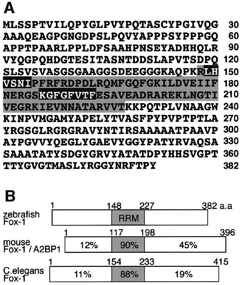
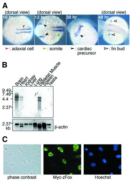
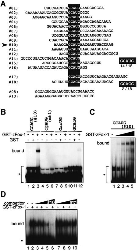
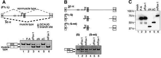
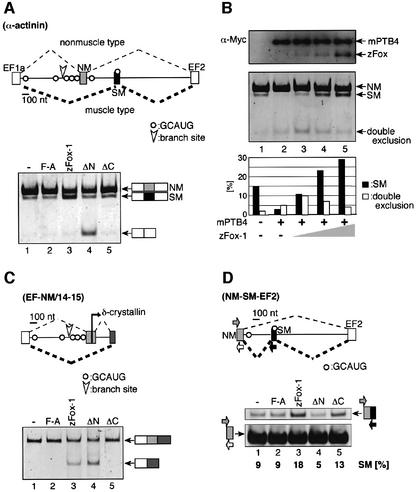
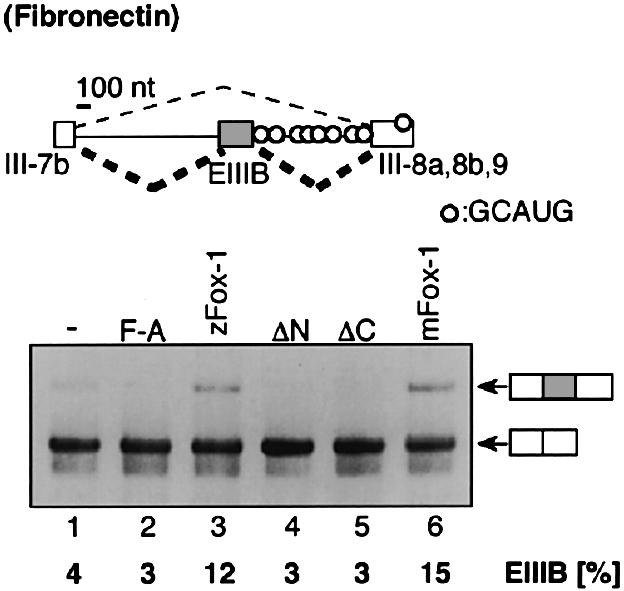
Alternative splicing is one of the central mechanisms that regulate eukaryotic gene expression. Here we report a tissue-specific RNA-binding protein, Fox-1, which regulates alternative splicing in vertebrates. Fox-1 bound specifically to a pentanucleotide GCAUG in vitro. In zebrafish and mouse, fox-1 is expressed in heart and skeletal muscles. As candidates for muscle-specific targets of Fox-1, we considered two genes, the human mitochondrial ATP synthase gamma-subunit gene (F1gamma) and the rat alpha-actinin gene, because their primary transcripts contain several copies of GCAUG. In transfection experiments, Fox-1 induced muscle-specific exon skipping of the F1gamma gene via binding to GCAUG sequences upstream of the regulated exon. Fox-1 also regulated mutually exclusive splicing of the alpha-actinin gene, antagonizing the repressive effect of polypyrimidine tract-binding protein (PTB). It has been reported that GCAUG is essential for the alternative splicing regulation of several genes including fibronectin. We found that Fox-1 promoted inclusion of the fibronectin EIIIB exon. Thus, we conclude that Fox-1 plays key roles in both positive and negative regulation of tissue-specific splicing via GCAUG.


Figures

None
Fig. 1. Identification of zebrafish and mouse Fox-1 proteins. (A) Amino acid sequence of zebrafish Fox-1. The RNA recognition motif (RRM) is shaded (Burd and Dreyfuss, 1994). The RNP2 (hexamer) and RNP1 (octamer) motifs are boxed in black. (B) Schematic representation of zebrafish, mouse and nematode Fox-1 proteins. The percentage identity of amino acid sequences is shown.
None
Fig. 2. Expression of zebrafish and mouse fox-1 genes. (A) Whole-mount in situ hybridization of zebrafish embryos using a fox-1 probe. A dorsal view of a 10 h embryo shows fox-1 expression in adaxial cells, precursor cells of slow muscle. A dorsal view of a 12 h embryo shows fox-1 expression in somites and bilateral presumptive heart cells. Expression in the developing heart as well as in somites is observed at 30 h. A dorsal view of a 48 h embryo shows expression in the finbud cells. (B) Northern blot analysis of mouse Fox-1 (upper) and β-actin (lower) using MessageMap Blot (Stratagene). (C) Nuclear localization of Fox-1 protein fused with a Myc tag was detected in CV-1 cells. Hoechst staining of the cells is shown in the right panel.
None
Fig. 3. Fox-1 protein binds specifically to GCAUG in vitro. (A) In the in vitro selection experiment, the sequences of 18 cDNA clones were aligned. The sequences GCAUG and GCACG are boxed. (B) Gel shift analyses of Fox-1 protein. No. 10 RNA (lanes 1–3) and mutant RNAs (lanes 4–12) were incubated with GST (lanes 2, 5, 8 and 11) or GST–zFox-1 (lanes 3, 6, 9 and 12). The position of unbound probe is shown by an asterisk. (C) Dose-dependent binding of Fox-1 protein to No. 10 RNA. Various amounts of GST–zFox-1 protein were incubated with the No. 10 RNA (0, 250, 500, 750 or 1000 ng, from left to right). (D) Competition experiments for GCAUG binding. No. 10 RNA was used as a probe. No.10 (lanes 3–6) or its mutant RNA (cgAUG) (lanes 7–10) was added as competitor (10-, 50-, 250-and 1250-fold excess compared with the 32P-labeled probe).
None
Fig. 4. Fox-1 induces muscle-specific splicing of the human mitochondrial ATP synthase F1γ gene via binding to GCAUG. (A) Transfection analyses with the hF1γL mini-gene. Exon 9 is skipped in a muscle-specific manner (Hayakawa et al., 2002). The positions of the branch site (arrowhead) and the GCAUG sequences (open circles) are shown in the schematic representation of the mini-gene. The mini-gene was transfected into L929 cells along with pCS2+ vector (Rupp et al., 1994) (lane 1), zFox-1 F190A (lane 2), zFox-1 (lane 3), zFox-1ΔN (lane 4), zFox-1ΔC (lane 5) or mouse Fox-1 (lane 6). The positions of non-muscle and muscle-type splicing products are indicated on the left. (B) Transfection analyses of the hF1γS mini-gene containing the Fox-1-binding sequence GCAUG (lanes 1–3) or the mutant sequence cgAUG (lanes 4–6) with pCS2+ MT vector (lanes 1 and 4), zFox-1 (lanes 2 and 5) or mouse Fox-1 (lanes 3 and 6). (C) Western blotting of the cell extracts to detect Fox-1 proteins: mock (lane 1), zFox-1 F190A (lane 2), zFox-1 (lane 3), zFox-1ΔN (lane 4), zFox-1ΔC (lane 5) and mouse Fox-1 (lane 6), expressed from the pCS2+ MT vector using the anti-Myc antibody.
None
Fig. 5. Muscle-specific splicing of the rat α-actinin gene is induced by Fox-1. (A) Transfection analyses of the rat α-actinin mini-gene (Southby et al., 1999; Suzuki et al., 2002). In the schematic representation, NM and SM represent non-muscle- and smooth muscle-specific exons, respectively. The branch site upstream of the NM exon and the GCAUG sequences are shown. The mini-gene was co-expressed in CV-1 cells along with pCS2+ MT vector (lane 1), zFox-1 F190A (lane 2), zFox-1 (lane 3), zFox-1ΔN (lane 4) or zFox-1ΔC (lane 5). The position of each mRNA product is indicated on the right. (B) Antagonism between the effects of Fox-1 and mouse PTB4. Transfection of the actinin mini-gene with pCS2+ MT vector (lane 1) or mouse PTB4 (lanes 2–5: 1.5 µg of the expression plasmid) and zFox-1 (lanes 3–5: 0.03, 0.09 and 0.27 µg, respectively, of the plasmid). The upper panel shows western blotting of the cell extracts to detect Fox-1 and PTB proteins expressed from the pCS2+ MT vector using the anti-Myc antibody. The splicing products from the actinin mini-gene were analyzed by RT–PCR (middle panel), and the amounts of SM mRNA (black bars) and EF1-EF2 mRNA (white bars) were expressed relative to the total amount of splicing products (lower panel). (C) Transfection analyses of the chimera construct, EF-NM/14–15, with pCS2+ MT vector (lane 1), zFox-1 F190A (lane 2), zFox-1 (lane 3), zFox-1ΔN (lane 4) or zFox-1ΔC (lane 5). The position of each mRNA product is shown schematically on the right. (D) Transfection analyses of the NM-SM-EF2 mini-gene with pCS2+ MT vector (lane 1), zFox-1 F190A (lane 2), zFox-1 (lane 3), zFox-1 ΔN (lane 4) or zFox-1 ΔC (lane 5). The primer sets are shown schematically (arrows). The fraction of SM exon inclusion (percentage) is shown at the bottom of each lane.
None
Fig. 6. Inclusion of the rat fibronectin EIIIB exon is promoted by Fox-1. The fibronectin 7iBi89 mini-gene (Huh and Hynes, 1993, 1994) was transfected into CV-1 cells with pCS2+ MT vector (lane 1), zFox-1 F190A (lane 2), zFox-1 (lane 3), zFox-1ΔN (lane 4), zFox-1ΔC (lane 5) or mouse Fox-1 (lane 6). The position of each mRNA product is shown on the right. The fraction of EIIIB exon inclusion (percentage) is shown at the bottom of each lane.

References

    1. Abe R., Sakashita,E., Yamamoto,K. and Sakamoto,H. (1996) Two different RNA binding activities for the AU-rich element and the poly(A) sequence of the mouse neuronal protein mHuC. Nucleic Acids Res., 24, 4895–4901. - PMC - PubMed
    1. Black D.L. (2000) Protein diversity from alternative splicing: a challenge for bioinformatics and post-genome biology. Cell, 103, 367–370. - PubMed
    1. Brudno M., Gelfand,M.S., Spengler,S., Zorn,M., Dubchak,I. and Conboy,J.G. (2001) Computational analysis of candidate intron regulatory elements for tissue-specific alternative pre-mRNA splicing. Nucleic Acids Res., 29, 2338–2348. - PMC - PubMed
    1. Burd C.G. and Dreyfuss,G. (1994) Conserved structures and diversity of functions of RNA-binding proteins. Science, 265, 615–621. - PubMed
    1. Charlet B.N., Logan,P., Singh,G. and Cooper,T.A. (2002) Dynamic antagonism between ETR-3 and PTB regulates cell type-specific alternative splicing. Mol. Cell, 9, 649–658. - PubMed

Publication types

Associated data