. 2003 Feb 17;22(4):913-24.
doi: 10.1093/emboj/cdg083.

Isolation of a U-insertion/deletion editing complex from Leishmania tarentolae mitochondria

Affiliations

Isolation of a U-insertion/deletion editing complex from Leishmania tarentolae mitochondria

Ruslan Aphasizhev et al. EMBO J. .

Abstract

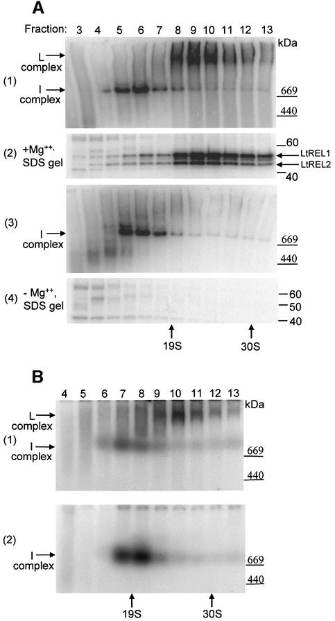
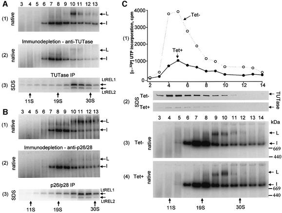
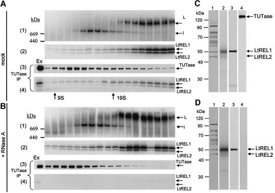
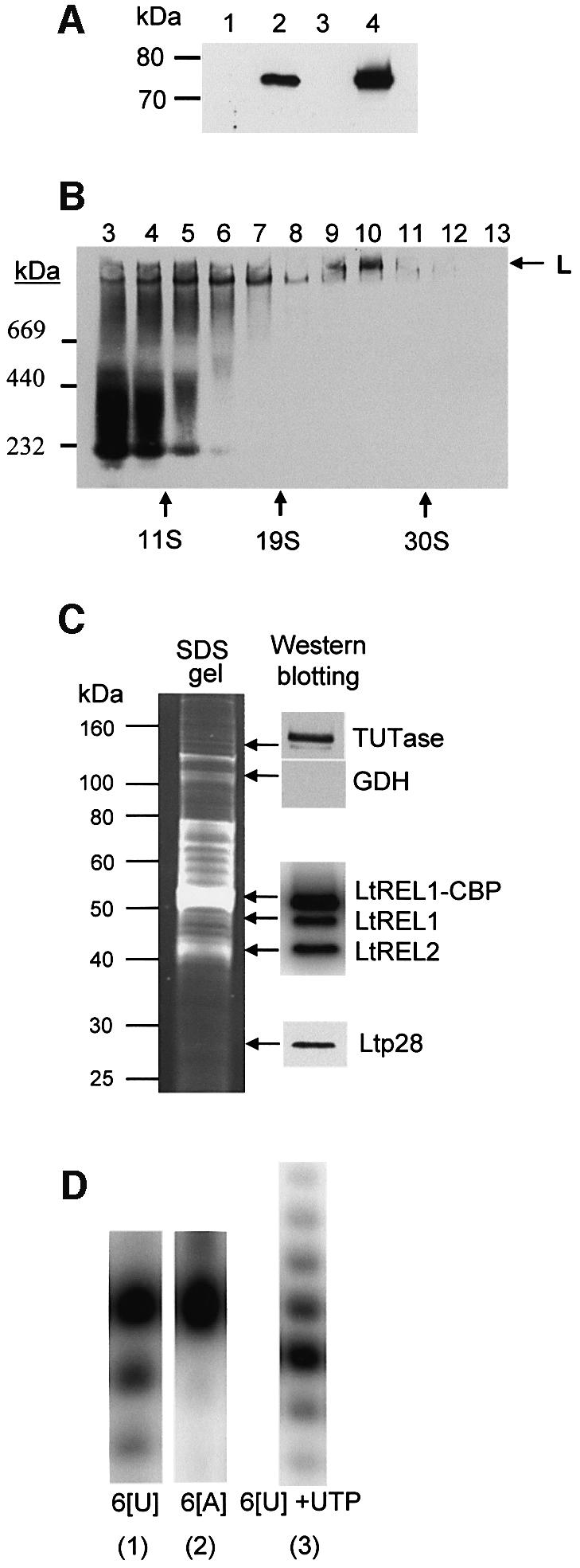
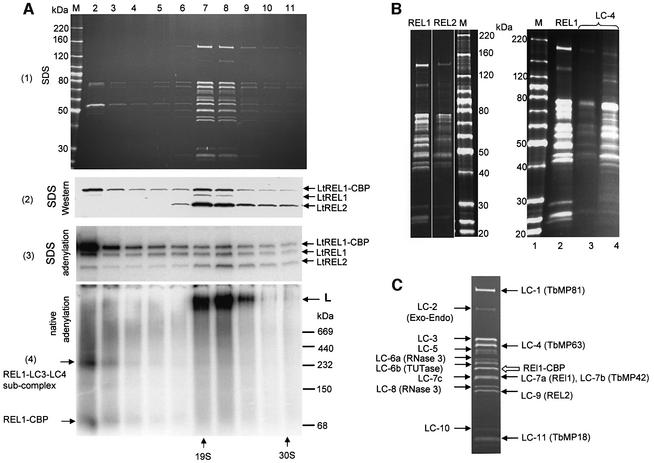
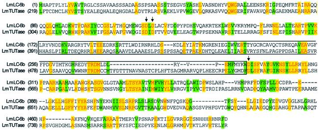
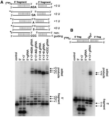
A multiprotein, high molecular weight complex active in both U-insertion and U-deletion as judged by a pre-cleaved RNA editing assay was isolated from mitochondrial extracts of Leishmania tarentolae by the tandem affinity purification (TAP) procedure, using three different TAP-tagged proteins of the complex. This editing- or E-complex consists of at least three protein-containing components interacting via RNA: the RNA ligase-containing L-complex, a 3' TUTase (terminal uridylyltransferase) and two RNA-binding proteins, Ltp26 and Ltp28. Thirteen approximately stoichiometric components were identified by mass spectrometric analysis of the core L-complex: two RNA ligases; homologs of the four Trypanosoma brucei editing proteins; and seven novel polypeptides, among which were two with RNase III, one with an AP endo/exonuclease and one with nucleotidyltransferase motifs. Three proteins have no similarities beyond kinetoplastids.


Figures

None
Fig. 1. Identification of the L-complex in mitochondrial extracts of L.tarentolae and T.brucei. The L-complex and the I-complex can be distinguished by labeling with [α-32P]ATP in the presence and absence of Mg2+. Mitochondrial S100 extracts were separated on a 10–30% glycerol gradient in an SW41 rotor for 20 h at 30 000 r.p.m. (ALeishmania tarentolae. Panel 1: gradient fractions were incubated with [α-32P]ATP in the presence of Mg2+ and separated on a 4–20% native gel. The locations of the L-complex and I-complex are indicated by arrows. Panel 2: same fractions as in 1, SDS gel. The adenylated LtREL1 and LtREL2 proteins are indicated. Panel 3: fractions incubated with [α-32P]ATP in the absence of Mg2+, 4–20% native gel. Panel 4: same fractions as in panel 3, SDS gel. (BTrypanosoma brucei. Panel 1: same conditions as panel 1 in (A). Panel 2: same conditions as panel 3 in (A).
None
Fig. 2. Interactions of the L-complex with 3′ TUTase and Ltp26/Ltp28 RNA-binding proteins. (A) Immunodepletion and co-immunoprecipitation of the L-complex with anti-TUTase antibody. Mitochondrial extract from L.tarentolae was fractionated on a 10–30% glycerol gradient for 20 h at 30 000 r.p.m. in an SW41 rotor, and two rounds of immunodepletion with affinity-purified antiserum against glutamate or antigen-purified polyclonal antibodies against recombinant TUTase were performed on each fraction. Panel 1: GDH immunodepletion control, native gel of adenylated fractions. The adenylated L-complex and I-complex bands are indicated. Panel 2: TUTase immunodepletion, native gel of adenylated fractions. Panel 3: IgG–Sepharose beads from the TUTase immunoprecipitation in panel 2 were incubated with [α-32P]ATP, washed and analyzed on a 10–20% SDS gel. The adenylated RNA ligase proteins are indicated. (B) Immunodepletion and co-immunoprecipitation of L-complex with antibodies against Ltp26 and Ltp28 RNA-binding proteins. Panel 1: control, native gel of adenylated gradient fractions after incubation with IgG beads saturated with pre-immune serum. Panel 2: native gel of Ltp26/Ltp28-immunodepleted adenylated gradient fractions. Panel 3: SDS gel of adenylated gradient fractions from IgG beads from panel 2. (C) Formation of L-complex in T.brucei procyclic cells is not affected by inhibition of TUTase expression. RNAi was induced with tetracycline for 3 days in procyclic T.brucei to down-regulate TUTase expression as described previously (Aphasizhev et al., 2002). Mitochondrial extracts were fractionated on 10–30% glycerol gradients for 20 h at 30 000 r.p.m. in an SW41 rotor. Panel 1: gradient fractions from uninduced and RNAi-induced cells were assayed for TUTase activity by incorporation of a [α-32P]UTP into a synthetic RNA substrate. Panel 2: TUTase western of SDS gels of gradient fractions from uninduced (Tet–) and RNAi-induced (Tet+) cells. Panels 3 and 4: native gels of adenylated gradient fractions from uninduced and RNAi-induced cells. The adenylated L-complex and I-complex bands are indicated by arrows.
None
Fig. 3. Interaction of L-complex with the 121 kDa 3′ TUTase is disrupted by RNase A treatment. Mitochondrial extracts were incubated without (A) or with (B) RNase A (0.1 mg/ml) and fractionated on 10–30% glycerol gradients in an SW 28 rotor for 40 h at 24 000 r.p.m. Panel 1: 8–16% Tris-glycine native gel of adenylated gradient fractions. Panel 2: SDS gel of adenylated gradient fractions from panel 1. Panel 3: western blotting detection of TUTase immunoprecipitated with anti-TUTase antibody and recovered from IgG beads. Panel 4: SDS gel of adenylated LtREL1 and LtREL2 proteins co-immunoprecipitated with anti-TUTase antibody and recovered from IgG beads. (C) The L-complex band from (A) was excised and the protein components eluted and separated in an SDS gel, which was silver stained. Lane 1, stained gel; lane 2, autoradiograph of adenylated LtREL1 and LtREL2; lane 3, western blot using anti-TbREL1 monoclonal P3C1-G2 antibody; lane 4, western blotting with anti-TUTase antibody. (D) The L-complex band from the RNase-treated extract in (B) was eluted as in (C). Lane 1, stained gel; lane 2, autoradiograph of adenylated LtREL1 and LtREL2; lane 3, western blot using anti-TbREL1 monoclonal P3C1-G2 antibody; lane 4, western blotting with anti-TUTase antibody.
None
Fig. 4. Expression of LtREL1–TAP fusion protein and affinity isolation of the L-complex. (A) Mitochondrial localization of LtREL1–TAP fusion protein. Total protein (10 µg) from wild-type cells (lane 1), transfected (lane 2) cells, the cytoplasmic fraction of transfected cells (lane 3), and 2 µg of protein from purified mitochondria of transfected cells (lane 4) were analyzed with the PAP reagent for the presence of protein A as a part of the TAP fusion protein. (B) The REL1–TAP fusion protein is incorporated into the L-complex. Mitochondrial S100 extract from transfected cells was fractionated on a 10–30% glycerol gradient in an SW41 rotor for 20 h at 30 000 r.p.m. Each fraction was separated on a 4–12% native gel, and the protein A fusion protein was detected with the PAP reagent. (C) Protein composition of the TAP-purified L-complex. The fraction eluted from the calmodulin column was analyzed for the presence of both LtREL1 and LtREL2 ligases by adenylation, and for TUTase and p28 by western analysis with the respective polyclonal antibodies. (D) Enzymatic activities of the TAP-purified complex. The same fraction as in (C) was analyzed for the presence of 3′–5′ U-specific exonuclease activity by incubating the calmodulin fraction with 5′-labeled synthetic RNA oligonucleotides with six uridine (6[U]) or adenosine (6[A]) residues at the 3′ end. 3′ TUTase activity was detected by including 100 µM UTP. Products were separated on 15% polyacrylamide–urea.
None
Fig. 5. Characterization of LtREL1–TAP associated proteins. (A) The TAP-purified fraction was separated further on a 10–30% glycerol gradient and each fraction was subjected to adenylation with [α-32P]ATP and analyzed by native and SDS gel electrophoresis. Panel 1: protein profile on an 8–16% SDS gel, Sypro Ruby staining. Panel 2: western analysis of RNA ligase proteins with polyclonal antibodies against both LtREL1 and LtREL2, SDS gel. Panel 3: adenylated proteins, SDS gel. The LtREL1–CBP and the two endogenous ligases are indicated. Panel 4: detection of adenylated L-complex sedimenting at ∼20S (arrow labeled L) and an ∼200 kDa LtREL1-containing subcomplex in fractions 2 and 3 (arrows on the left), native gel. (B) Comparison of major polypeptides from LtREL2–TAP and LmLC-4–TAP complex with those from the LtREL1–TAP complex. Left panel: REL1, gradient-purified LtREL1–TAP complex; REL2, gradient-purified LtREL2–TAP complex; M, molecular weight markers. Right panel: M, molecular weight markers; REL1, gradient-purified LtREL1–TAP complex; LC-4, LmLC-4–TAP complex. Lane 4 is the material after the calmodulin elution and lane 3 is the concentrated 20–25S gradient fractions. Sypro Ruby-stained, SDS gel. (C) Identification of individual bands of L-complex by mass spectroscopy and database analysis. The lane shown is the Sypro Ruby-stained fraction 8 from (A). Novel proteins corresponding to L.major unidentified open reading frames are indicated on the left, and proteins homologous to known T.brucei proteins on the right. See Table I for details of proteins.
None
Fig. 6. Partial alignment of the 57 kDa putative 3′ TUTase with the 121 kDa 3′ TUTase from L.major. The three aspartates of the nucleotidyltransferase catalytic triad are indicated by arrows, and the TUTase-specific insertion between D2 and D3 is indicated by boxing. Alignment performed by AlignX in Vector NTI.
None
Fig. 7. In vitro gRNA-directed U-insertion and U-deletion activities of LtREL1–TAP purified material. (A) U-insertion in vitro activity of the LtREL1–TAP purified material. Fraction 8 from Figure 5 was incubated with pre-annealed RNA substrates for the pre-cleaved insertion assay in the presence of 200 µM UTP and 20 µM ATP, and products were separated on a 15% acrylamide–urea gel. The bridged RNA substrates are shown schematically above. Guiding nucleotides are indicated in bold. ‘0’ indicates no guiding nucleotide. ‘CCC’ indicates three non-guiding C nucleotides. The locations of the ligated edited products are indicated, as are the 5′ fragments with 1–3 Us added. Control, no protein added. (B) U-deletion in vitro activity. The same fraction as in (A) was incubated with RNA substrates for pre-cleaved deletion assay. The bridged substrate which should guide the deletion of two Us is shown schematically.

References

    1. Adler B.K., Harris,M.E., Bertrand,K.I. and Hajduk,S.L. (1991) Modification of Trypanosoma brucei mitochondrial rRNA by posttranscriptional 3′ polyuridine tail formation. Mol. Cell. Biol., 11, 5878–5884. - PMC - PubMed
    1. Allen T.E., Heidmann,S., Reed,R., Myler,P.J., Goringer,H.U. and Stuart,K.D. (1998) Association of guide RNA binding protein gBP21 with active RNA editing complexes in Trypanosoma brucei. Mol. Cell. Biol., 18, 6014–6022. - PMC - PubMed
    1. Aphasizhev R. and Simpson,L. (2001) Isolation and characterization of a U-specific 3′–5′ exonuclease from mitochondria of Leishmania tarentolae. J. Biol. Chem., 276, 21280–21284. - PubMed
    1. Aphasizhev R., Sbicego,S., Peris,M., Jang,S.H., Aphasizheva,I., Simpson,A.M., Rivlin,A. and Simpson,L. (2002) Trypanosome mitochondrial 3′ terminal uridylyl transferase (TUTase): the key enzyme in U-insertion/deletion RNA editing. Cell, 108, 637–648. - PubMed
    1. Aphasizhev R., Aphasizheva,I., Nelson,R.E. and Simpson,L. (2003) A 100 kDa complex of two RNA-binding proteins from mitochondria of Leishmania tarentolae catalyzes RNA annealing and interacts with several editing components. RNA, 9, 62–76. - PMC - PubMed

Publication types

Associated data