. 2003 Feb 17;22(4):954-63.
doi: 10.1093/emboj/cdg091.

E1 initiator DNA binding specificity is unmasked by selective inhibition of non-specific DNA binding

Affiliations

E1 initiator DNA binding specificity is unmasked by selective inhibition of non-specific DNA binding

Arne Stenlund. EMBO J. .

Abstract

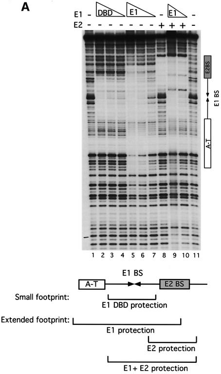
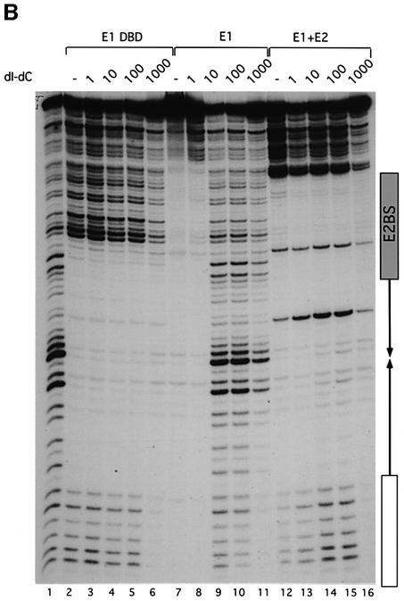
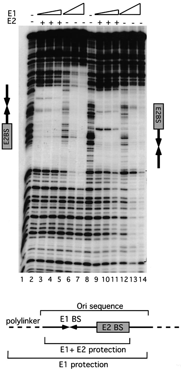
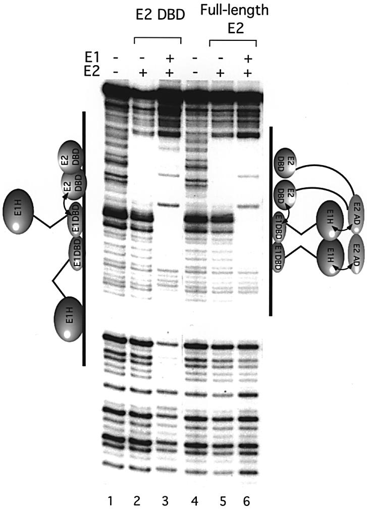
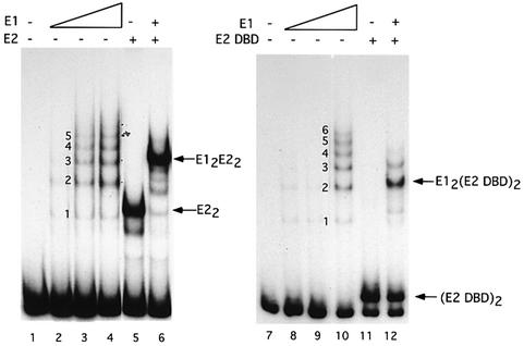
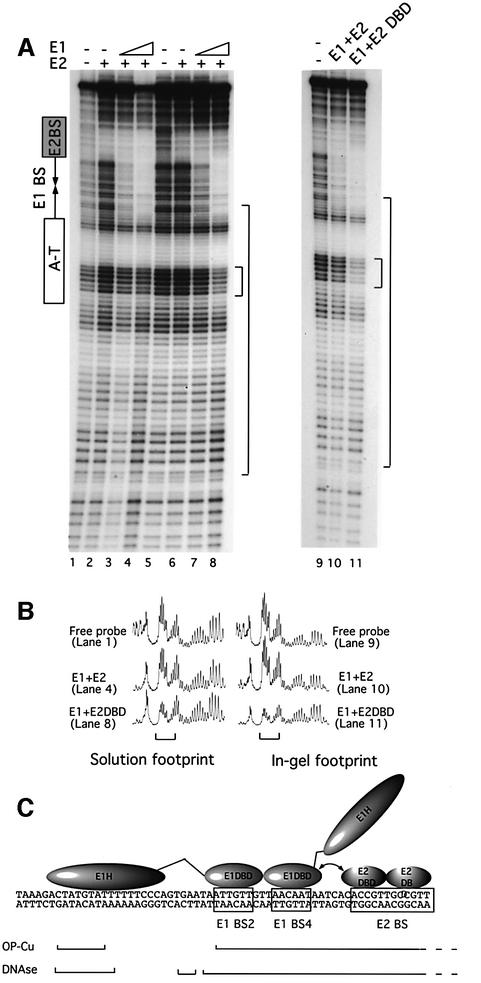
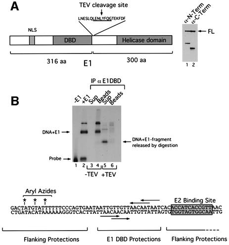
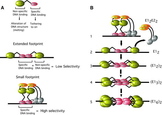
Initiator proteins are critical components of the DNA replication machinery and mark the site of initiation. This activity probably requires highly selective DNA binding; however, many initiators display modest specificity in vitro. We demonstrate that low specificity of the papillomavirus E1 initiator results from the presence of a non-specific DNA-binding activity, involved in melting, which masks the specificity intrinsic to the E1 DNA-binding domain. The viral factor E2 restores specificity through a physical interaction with E1 that suppresses non-specific binding. We propose that this arrangement, where one DNA-binding activity tethers the initiator to ori while another alters DNA structure, is a characteristic of other viral and cellular initiator proteins. This arrangement would provide an explanation for the low selectivity observed for DNA binding by initiator proteins.


Figures

None
Fig. 1. (A) E1 and the E1 DBD bind the ori differently. DNase I footprinting was carried out on the top strand of the minimal ori using the E1 DBD (8, 4 and 2 ng, lanes 2–4), full-length E1 (20, 10 and 5 ng, lanes 5–7), full-length E2 (1 ng, lane 8) and full-length E1 (10 and 5 ng) and E2 (1 ng) together (lanes 9 and 10). Shown below is the extent of protection generated by the different proteins relative to the AT-rich region (A-T), the E1-binding sites (E1 BS) and the E2-binding site (E2 BS). (B) The E1 DBD binds DNA with a high degree of selectivity while full-length E1 binds with low selectivity. DNase footprinting was carried out on the top strand of the minimal ori using the E1 DBD (8 ng, lanes 2–6), full-length E1 (20 ng, lanes 7–11) and full-length E1 and E2 (5 and 1 ng, respectively, lanes 12–16). Competitor DNA, poly(dI–dC), at the indicated concentrations was mixed with the probe followed by the addition of protein. In lanes 1, 2, 7 and 12, no competitor DNA was added.
None
Fig. 2. The E1 footprint extends symmetrically over two helical turns of flanking sequence. An ori fragment spanning the E1- and E2-binding sites, but lacking the AT-rich region, was inserted into the polylinker of pUC19 in two orientations to provide two different contexts of flanking sequences. Probes were generated from both orientations, and DNase footprinting was performed with E1 (10, 5 and 2.5 ng) and E2 (1 ng) in combination (lanes 2–4 and lanes 9–11), or with E1 alone (5, 10 and 20 ng, lanes 5–7 and lanes 12–14). Below is shown schematically the extent of the protections relative to the elements in the ori.
None
Fig. 3. E1 generates the small footprint together with E2, but the extended footprint with the E2 DBD. DNase footprinting was performed on the top strand of the wild-type ori using E2 alone (1 ng, lane 5), the E2 DBD alone (0.5 ng, lane 2), E1 and E2 (5 and 1 ng, respectively, lane 6), and E1 and the E2 DBD (5 and 0.5 ng, respectively, lane 3).
None
Fig. 4. Formation of E12E22 and E12(E2 DBD)2 complexes. EMSA was performed using full-length E1 alone (0.5, 1 and 2 ng, lanes 2–4 and lanes 8–10) and E1 (0.5 ng) in the presence of E2 (0.1 ng, lane 6) or the E2 DBD (0.05 ng, lane 12). Lanes 5 and 11 contained 0.1 ng of E2 and 0.05 ng of the E2 DBD, respectively. Lanes 1 and 7 contained probe alone. The ladders generated by E1 alone in lanes 3 and 10 serve as markers. The number of molecules in each complex is indicated.
None
Fig. 5. In-gel OP-Cu footprinting of E12E22 and E12(E2 DBD)2 complexes. (A) Solution OP-Cu footprints were generated with E1 and E2 (lanes 1–4) or E1 and the E2 DBD (lanes 5–8). Two quantities of full-length E1, 25 and 50 ng, in the presence of 5 ng of E2 (lanes 3 and 4), or in the presence of 2.5 ng of the E2 DBD (lanes 7 and 8) were bound. Solution footprints with E2 and the E2 DBD alone (5 and 2.5 ng, respectively) are shown in lanes 2 and 6. In-gel OP-Cu footprints were performed on free probe (lane 9) and with complexes formed by E1 and E2 (lane 10) and complexes formed by E1 and the E2 DBD (lane 11). The large bracket indicates the section of the gels that was quantitated and is shown in (B). (B) The gels were exposed to a Fuji imaging plate and the profiles for six selected lanes are shown below. The small bracket indicates the flanking sequences that are protected in the E12(E2 DBD)2 complex. (C) A schematic summary of the DNase I and OP-Cu protections observed on the top strand for the E12(E2 DBD)2 complex in relation to the position of the E1- and E2-binding sites.
None
Fig. 6. Site-specific UV cross-linking demonstrates that the E1 helicase domain binds to the AT-rich region. (A) An E1 protein engineered with a TEV protease cleavage site inserted between the DBD and the helicase domain was expressed, purified and used for UV cross-linking experiments. Digestion of this protein with TEV protease generates two fragments, an N-terminal fragment of 35 kDa and a C-terminal fragment of 30 kDa, which can be detected by western blotting using monoclonal antibodies directed against the N- (lane 1) and C-terminus (lane 2) of the E1 protein. (B) The probe for UV cross-linking was generated by modifying an oligonucleotide with three phosphorothioate linkages with azidophencyl bromide to generate aryl azides in the flanking sequence upstream (left) of the E1-binding sites. An ori probe was generated by PCR using the modified oligonucleotide and a 32P-labeled primer from the other strand. This probe was used for UV cross-linking to E1-TEV. After UV irradiation, the level of cross- linking in the absence or presence of E1 was assessed by SDS–PAGE (lanes 1 and 2). The UV-irradiated sample was purified by ion exchange chromatography and immunoprecipitated with a polyclonal antiserum against the E1 DBD (lanes 4–6). After the immunoprecipitation, the sample was divided in two, and incubated with (lanes 5 and 6) or without (lanes 3 and 4) TEV protease. Following digestion, the protein G beads (lanes 4 and 6) and the supernatants (lanes 3 and 5) were separated and analyzed by SDS–PAGE.
None
Fig. 7. (A) A summary of the dual DNA-binding activities in E1 and the consequences for DNA binding specificity. The E1 initiator encodes two distinct DNA-binding activities. A highly specific DNA-binding activity is present in the E1 DBD. This activity is responsible for recognition and tethering of E1 to the ori. The second DNA-binding activity is present in the helicase domain of E1, binds DNA with low specificity and is required for melting of the sequences flanking the E1-binding site. In the absence of E2, the non-specific DNA-binding activity masks the specificity of the E1 DBD, resulting in a net low selectivity for E1 DNA binding. In the presence of E2, however, the interaction between the E2 AD and the E1 helicase domain (E1H) inactivates the non-specific DNA-binding activity, resulting in unmasking of the intrinsic specificity of the E1 DBD. The extended footprint forms by binding of the E1 DBD with high specificity to the E1-binding sites, and simultaneous binding of the E1 helicase domain with low specificity to the flanking sequences. In the small footprint, DNA contacts are generated by the E1 DBD only. (B) A model for the assembly of E1 monomers to form a double hexamer. See text for details.

Similar articles

Cited by

References

    1. Aurora R. and Herr,W. (1992) Segments of the POU domain influence one another’s DNA binding specificity. Mol. Cell. Biol., 12, 455–467. - PMC - PubMed
    1. Bell S.P. (2002) The origin recognition complex: from simple origins to complex functions. Genes Dev., 16, 659–672. - PubMed
    1. Bell S.P. and Stillman,B. (1992) ATP-dependent recognition of eukaryotic origins of DNA replication by a multiprotein complex. Nature, 357, 128–134. - PubMed
    1. Berg M. and Stenlund,A. (1997) Functional interactions between papillomavirus E1 and E2 proteins. J. Virol., 71, 3853–3863. - PMC - PubMed
    1. Chen G. and Stenlund,A. (1998) Characterization of the DNA-binding domain of the bovine papillomavirus replication initiator E1. J. Virol., 72, 2567–2576. - PMC - PubMed

Publication types