. 2003 Feb 17;22(4):975-86.
doi: 10.1093/emboj/cdg082.

p53 is a chromatin accessibility factor for nucleotide excision repair of DNA damage

Affiliations

p53 is a chromatin accessibility factor for nucleotide excision repair of DNA damage

Carlos P Rubbi et al. EMBO J. .

Abstract

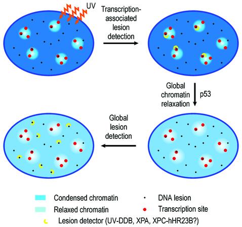
One of the longest standing problems in DNA repair is how cells relax chromatin in order to make DNA lesions accessible for global nucleotide excision repair (NER). Since chromatin has to be relaxed for efficient lesion detection, the key question is whether chromatin relaxation precedes lesion detection or vice versa. Chromatin accessibility factors have been proposed but not yet identified. Here we show that p53 acts as a chromatin accessibility factor, mediating UV-induced global chromatin relaxation. Using localized subnuclear UV irradiation, we demonstrate that chromatin relaxation is extended over the whole nucleus and that this process requires p53. We show that the sequence for initiation of global NER is as follows: transcription-associated lesion detection; p53-mediated global chromatin relaxation; and global lesion detection. The tumour suppressor p53 is crucial for genomic stability, a role partially explained by its pro-apoptotic capacity. We demonstrate here that p53 is also a fundamental component of DNA repair, playing a direct role in rectifying DNA damage.


Figures

None
Fig. 1. Antibody blocking of p53 inhibits UDS. Human NDFs were microinjected in the nucleus with an anti-p53 monoclonal antibody (DO-1) (A), an anti-XPA antibody (B) or purified mouse IgG (C). (D) Non-irradiated control. After UV irradiation at 20 J/m2, cells were assayed for UDS. Arrows indicate cells that received the micro injections. (A′–D′) are reference images of Hoechst 33258 staining.
None
Fig. 2. Trichostatin A overcomes the effect of p53 deficiency in NER. (A–D) TSA preferentially enhances NER in p53-deficient cells. (A and B) UDS of NDF incubated for 3 h in [3H]thymidine following UV irradiation either untreated (A) or pre-incubated for 20 h with 200 ng/ml TSA (B). (C and D) The same experiment with 041 p53-deficient fibroblasts, either untreated (C) or pre-incubated with TSA (D). (EG) Human NDFs were micro injected with anti-p53, anti-XPA or control antibodies exactly as in Figure 1, except that 200 ng/ml TSA was added to cultures 20 h prior to UV irradiation. Figure labels are as in Figure 1.
None
Fig. 3. UV-induced, p53-mediated global chromatin relaxation. (A) Micrococcal nuclease sensitivity as a validation of the HCl/AO assay, confirming the effect of UV irradiation and TSA on chromatin. (B) DNA denaturation sensitivity measured by the HCl/AO assay applied to human NDFs, p53-null 041 fibroblasts and XPE fibroblasts. Points indicate the extent of chromatin relaxation as the fraction of dsDNA fluorescence (green). Inserts show red/green AO images and pseudo-colour (dsDNA fraction) images. UV irradiation (4 J/m2) was assayed after 1 h, and TSA (200 ng/ml) was applied for 1 h. (C) Cells grown on Isopore filters, ‘back-irradiated’ with UV light, labelled with an anti-CPD antibody and counterstained with Hoechst 33258. (D and D′) Combination of the techniques used in (B) and (C). (E) Human NDFs and 041 fibroblasts treated as in (B), with or without the addition of 20 µg/ml α-amanitin 15 min prior to UV irradiation, and maintained throughout.
None
Fig. 4. p53 is required for UV-mediated histone acetylation. Human NDFs and p53-null 041 fibroblasts (A), and HCT116 human colon carcinoma cells either expressing wild-type p53 or p53–/– (B) were assayed by western blot for total histone H3 and Lys9-acetylated histone H3 (AcH3) at different times following UV irradiation at 4 J/m2. (C–H) Confocal images of cells fluorescently labelled for Lys9-acetylated histone H3. (C and D) Untreated NDFs and NDFs 4 h post-UV irradiation, respectively. (F and G) The same as (C and D) for 041 fibroblasts. NDFs microinjected in the nucleus with either purified mouse IgG (E) or an anti-p53 antibody (DO-1) (H), both UV irradiated and labelled for AcH3.
None
Fig. 5. p53 co-localizes with sites of NER, while for p300 this co-localization is p53 dependent. Single confocal sections of nuclei of human NDFs double labelled for p53 or p300 (green) and NER sites detected through transient ssDNA (red), 20 min after UV irradiation at 20 J/m2. (A and B) NDFs labelled for NER sites and for p53 and p300, respectively. (C) p53-null 041 fibroblasts labelled for NER sites and p300. All bars are 5 µm. (A′), (B′) and (C′) are line profiles of red and green images taken at the positions indicated by lines in the confocal sections. (A”), (B”) and (C”) are plots of Pearson’s correlation coefficients at a range of x-axis shifts of green images with respect to red; black and red lines correspond to plots of whole and thresholded images, respectively. (D and E) NDFs microinjected with anti-p300 purified polyclonal IgG or with non-specific IgG, respectively, and assayed for UDS as in Figure 1. (D′ and E′) Hoechst images showing the positions of nuclei. Arrows indicate injected cells.
None
Fig. 6. Proposed mechanism. Chromatin relaxation events in the initiation of GGR suggested from the results in Figures 1–3. See text.

Similar articles

Cited by

References

    1. Ait-Si-Ali S. et al. (2000) CBP/p300 histone acetyl-transferase activity is important for the G1/S transition. Oncogene, 19, 2430–2437. - PubMed
    1. Benhamou S. and Sarasin,A. (2000) Variability in nucleotide excision repair and cancer risk: a review. Mutat. Res., 462, 149–158. - PubMed
    1. Bunz F., Dutriaux,A., Lengauer,C., Waldman,T., Zhou,S., Brown,J.P., Sedivy,J.M., Kinzler,K.W. and Vogelstein,B. (1998) Requirement for p53 and p21 to sustain G2 arrest after DNA damage. Science, 282, 1497–1501. - PubMed
    1. Christians F.C. and Hanawalt,P.C. (1992) Inhibition of transcription and strand-specific DNA repair by α-amanitin in Chinese hamster ovary cells. Mutat. Res., 274, 93–101. - PubMed
    1. Cleaver J.E. and Thomas,G.H. (1981) Measurement of unscheduled DNA synthesis by autoradiography. In Friedberg,E.C. and Hanawalt,P.C. (eds), DNA Repair: A Laboratory Manual of Research Procedures. Vol I, part B. Marcel Dekker, New York, NY, pp. 277–287.

Publication types