. 2003 Feb;41(2):717-22.
doi: 10.1128/JCM.41.2.717-722.2003.

Comparison of PCR assays for detection of the agent of human granulocytic ehrlichiosis, Anaplasma phagocytophilum

Affiliations

Comparison of PCR assays for detection of the agent of human granulocytic ehrlichiosis, Anaplasma phagocytophilum

Robert F Massung et al. J Clin Microbiol. 2003 Feb.

Abstract

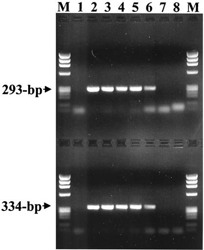
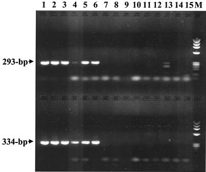
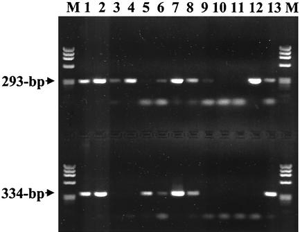
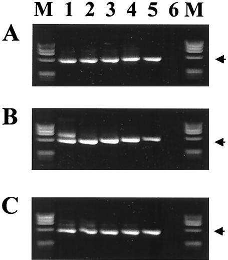
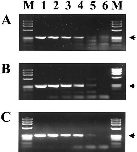
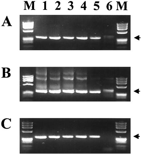
Human granulocytic ehrlichiosis is an emerging infectious disease in the United States and Europe, and PCR methods have been shown to be effective for the diagnosis of acute infections. Numerous PCR assays and primer sets have been reported in the literature. The analytical sensitivities (limits of detection) of 13 published PCR primer sets were compared using DNA extracted from serial dilutions of Anaplasma phagocytophilum-infected HL-60 cells. The specificity of the assays that were able to detect <or=2.5 infected cells was tested by the use of template DNA extracted from Ehrlichia chaffeensis, Rickettsia rickettsii, and Bartonella henselae. The assays with the lowest limits of detection were shown to be a nested assay that amplifies the 16S rRNA gene (primer pairs ge3a-ge10 [primary] and ge9-ge3 [nested]; detects 0.25 infected cell), a direct assay that amplifies the major surface protein gene msp2 (primer pair msp2-3f-msp2-3r; detects 0.25 infected cell), and a direct assay that amplifies the 16S rRNA gene (primer pair ehr521-ehr790; detects 0.25 infected cell). The specificity and limit of detection of the MSP2 and 16S rRNA direct assays were further tested by use of A. phagocytophilum template DNA from both North America and Europe and from human, tick, white-footed mouse, equine, deer, bovine, and wood rat samples and of template DNA from closely related species (Anaplasma marginale, the white-tailed deer agent, and additional E. chaffeensis-positive samples). Three manufacturers' PCR kits were tested and showed distinct variations in the limit of detection, specificity, and nonspecific background amplification. The importance of these results for the molecular diagnosis of human granulocytic ehrlichiosis is discussed.


Figures

FIG. 1.
FIG. 1.
Limit of detection of PCR assays for A. phagocytophilum strain USG3 DNA with primer pairs ehr521-ehr790 (upper lanes) and msp2-3f-msp2-3r (lower lanes). Lanes 1 show amplification products from the DNA extracted from uninfected human blood as negative controls. Lanes 2 through 8 represent amplifications from a 10-fold dilution series ranging from 5 × 10⁵ infected cells/ml to 0.5 infected cell/ml. Phage ϕX174 DNA digested with HaeIII was run in the lanes labeled M. The sizes of the amplified products are indicated to the left.
FIG. 2.
FIG. 2.
PCR assay testing on DNA extracted from human EDTA blood samples with primer pairs ehr521-ehr790 (upper lanes) and msp2-3f-msp2-3r (lower lanes). Lanes 1 through 6 show amplification products from samples previously confirmed as A. phagocytophilum positive by 16S-rRNA-nested PCR. Lanes 7 through 11 are amplification products from samples previously shown to be negative for A. phagocytophilum and E. chaffeensis. Lanes 12 through 15 show amplification products from samples previously confirmed as E. chaffeensis positive. Phage ϕX174 DNA digested with HaeIII was run in the lane labeled M. The sizes of the amplified products are indicated to the left.
FIG. 3.
FIG. 3.
PCR assay testing on DNA extracted from veterinary, tick, and cultured bacterial samples with primer pairs ehr521-ehr790 (upper lanes) and msp2-3f-msp2-3r (lower lanes). Samples represented are E. equi (now A. phagocytophilum) from an experimentally infected horse (lane 1), E. phagocytophila (now A. phagocytophilum) from a Swedish cow (lane 2), white-tailed deer agent from white-tailed deer (lanes 3 and 4), A. phagocytophilum-positive I. scapularis ticks from Connecticut (lanes 5 through 7), E. equi (now A. phagocytophilum) from a wood rat from California (lane 8), E. chaffeensis Arkansas strain (lane 9), B. henselae Houston-1 strain (lane 10), R. rickettsii Sheila Smith strain (lane 11), A. marginale (lane 12), and A. phagocytophilum USG3 strain (lane 13). Phage ϕX174 DNA digested with HaeIII was run in the lanes labeled M. The sizes of the amplified products are indicated to the left.
FIG. 4.
FIG. 4.
Comparison of the 16S-rRNA-nested-PCR assay with primer pairs ge3a-ge10 and ge9-ge2 with commercially available kits from three manufacturers: (A) PerkinElmer GeneAmp PCR reagent kit with AmpliTaq DNA polymerase; (B) Amersham Pharmacia Ready-To-Go PCR beads; (C) Qiagen Taq PCR Master Mix kit. Lanes 1 through 5 represent amplifications from a 10-fold dilution series ranging from 5 × 10⁵ A. phagocytophilum-infected cells/ml to 50 infected cells/ml, and lane 6 contains DNA from uninfected human blood as a negative control. Phage ϕX174 DNA digested with HaeIII was run in the lanes labeled M. The arrows indicate the position of the 546-bp amplified products.
FIG. 5.
FIG. 5.
Comparison of the 16S rRNA assay with primer pair ehr521-ehr790 by using commercially available kits from three manufacturers: (A) PerkinElmer GeneAmp PCR reagent kit with AmpliTaq DNA polymerase; (B) Amersham Pharmacia Ready-To-Go PCR beads; (C) Qiagen Taq PCR Master Mix kit. Lanes are as described in the legend to Fig. 4. The arrows indicate the position of the 293-bp amplified products.
FIG. 6.
FIG. 6.
Comparison of the msp2 gene PCR assay using commercially available kits from three manufacturers: (A) PerkinElmer GeneAmp PCR reagent kit with AmpliTaq DNA polymerase; (B) Amersham Pharmacia Ready-To-Go PCR beads; (C) Qiagen Taq PCR Master Mix kit. Lanes are as described in the legend to Fig. 4. The arrows indicate the position of the 334-bp amplified products.

References

    1. Aguero-Rosenfeld, M. E., F. Kalantarpour, M. Baluch, H. W. Horowitz, D. F. McKenna, J. T. Raffalli, T.-Z. Hsieh, J. Wu, J. S. Dumler, and G. P. Wormser. 2000. Serology of culture-confirmed cases of human granulocytic ehrlichiosis. J. Clin. Microbiol. 38:635-638. - PMC - PubMed
    1. Anderson, B. E., J. E. Dawson, D. C. Jones, and K. H. Wilson. 1991. Ehrlichia chaffeensis, a new species associated with human ehrlichiosis. J. Clin. Microbiol. 29:2838-2842. - PMC - PubMed
    1. Bakken, J. S., J. S. Dumler, S.-M. Chen, M. R. Eckman, L. L. Van Etta, and D. H. Walker. 1994. Human granulocytic ehrlichiosis in the upper Midwest United States. A new species emerging? JAMA 272:212-218. - PubMed
    1. Birtles, R. J., T. G. Harrison, and D. H. Molyneux. 1994. Grahamella in small woodland mammals in the U.K.: isolation, prevalence and host specificity. Ann. Trop. Med. Parasitol. 88:317-327. - PubMed
    1. Brouqui, P., and D. Raoult. 1992. In vitro susceptibility of the newly recognized agent of ehrlichiosis in humans, Ehrlichia chaffeensis. Antimicrob. Agents Chemother. 36:2799-2803. - PMC - PubMed

MeSH terms

LinkOut - more resources