. 2003 Feb 1;23(3):777-87.
doi: 10.1523/JNEUROSCI.23-03-00777.2003.

An extranuclear locus of cAMP-dependent protein kinase action is necessary and sufficient for promotion of spiral ganglion neuronal survival by cAMP

Affiliations

An extranuclear locus of cAMP-dependent protein kinase action is necessary and sufficient for promotion of spiral ganglion neuronal survival by cAMP

Jinwoong Bok et al. J Neurosci. .

Abstract

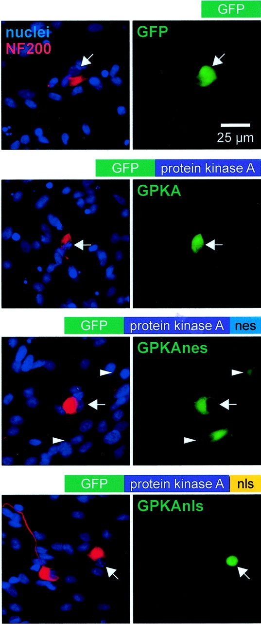
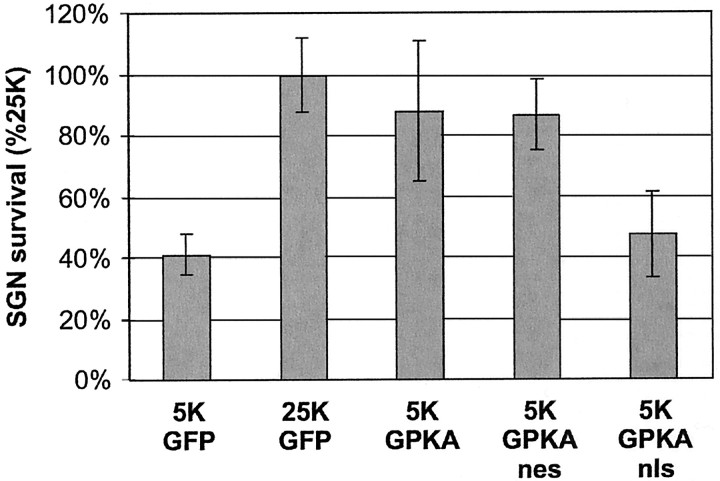
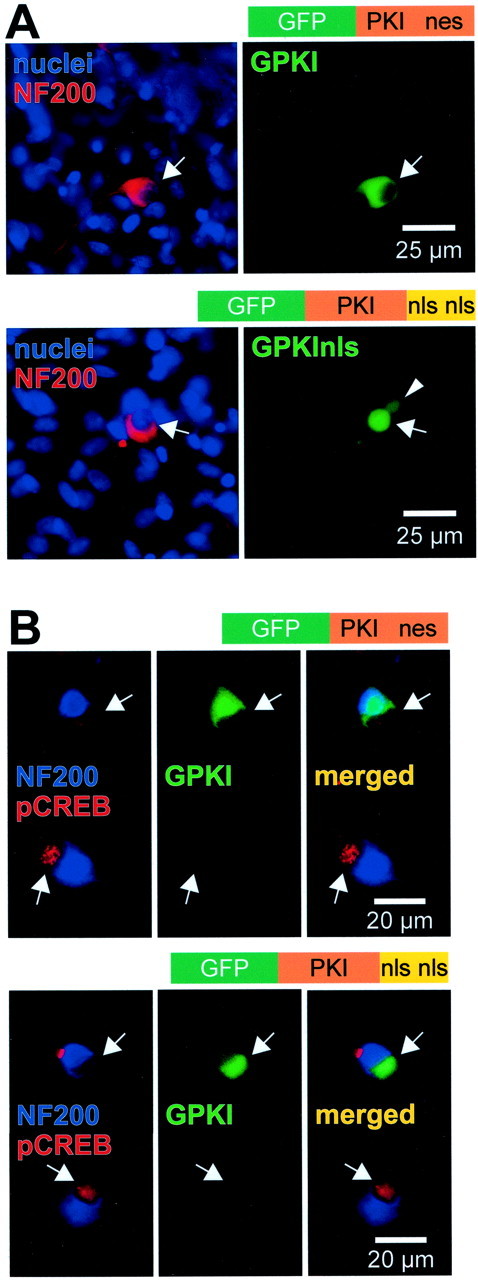
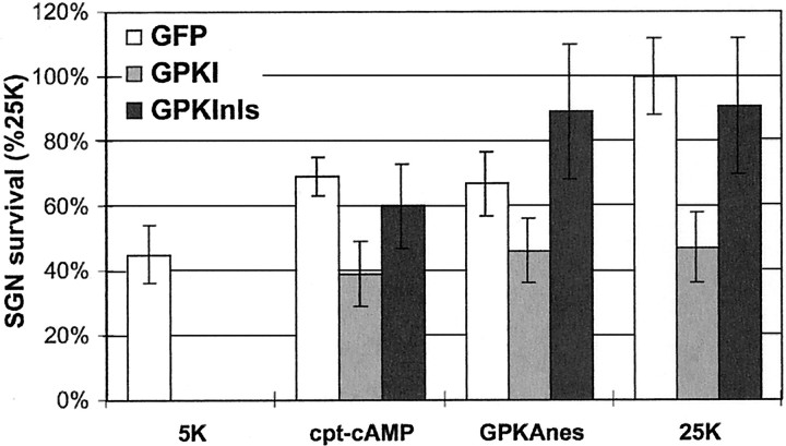
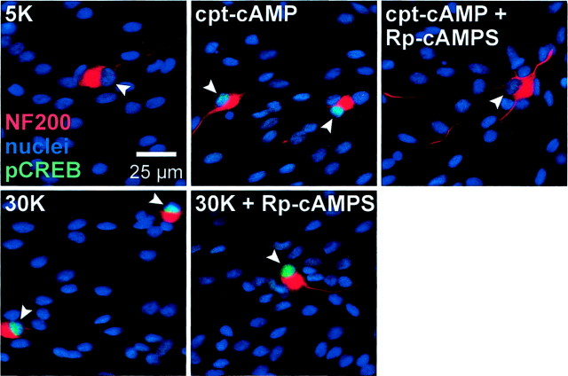
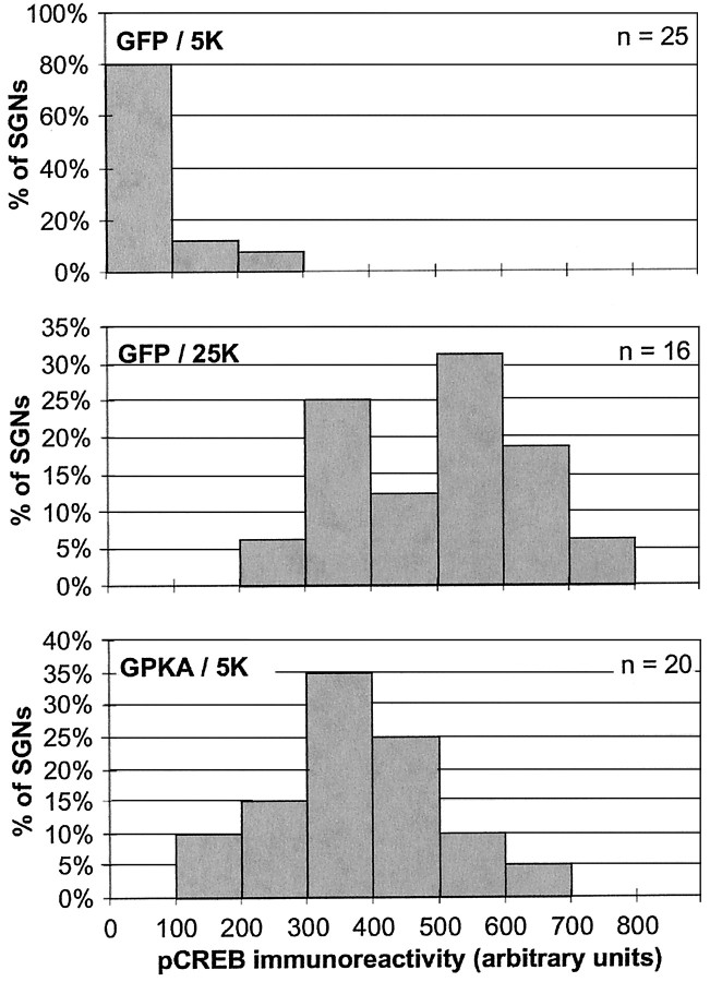
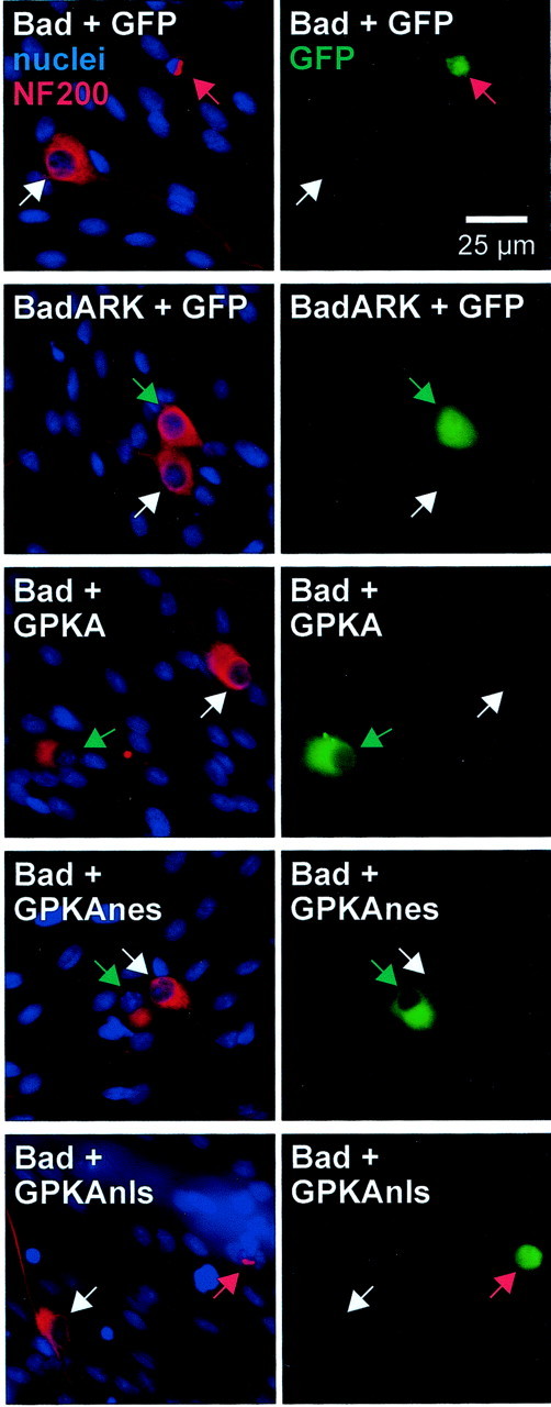
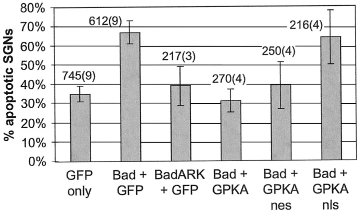
We showed previously that cAMP is a survival-promoting stimulus for cultured postnatal rat spiral ganglion neurons (SGNs) and that depolarization promotes SGN survival in part via recruitment of cAMP signaling. We here investigate the subcellular locus of cAMP prosurvival signaling. Transfection of GPKI, a green fluorescent protein (GFP)-tagged cAMP-dependent protein kinase (PKA) inhibitor, inhibits the ability of the permeant cAMP analog cpt-cAMP [8-(4-chlorophenylthio)-cAMP] to promote survival, indicating that PKA activity is necessary. Transfection of GFP-tagged PKA (GPKA) is sufficient to promote SGN survival, but restriction of GPKA to the nucleus by addition of a nuclear localization signal (GPKAnls) almost completely abrogates its prosurvival effect. In contrast, GPKA targeted to the extranuclear cytoplasm by addition of a nuclear export signal (GPKAnes) promotes SGN survival as effectively as does GPKA. Moreover, GPKI targeted to the nucleus lacks inhibitory effect on SGN survival attributable to cpt-cAMP or depolarization. These data indicate an extranuclear target of PKA for promotion of neuronal survival. Consistent with this, we find that dominant-inhibitory CREB mutants inhibit the prosurvival effect of depolarization but not that of cpt-cAMP. SGN survival is compromised by overexpression of the proapoptotic regulator Bad, previously shown to be phosphorylated in the cytoplasm by PKA. This Bad-induced apoptosis is prevented by cpt-cAMP or by cotransfection of GPKA or of GPKAnes but not of GPKAnls. Thus, cAMP prevents SGN death through a cytoplasmic as opposed to nuclear action, and inactivation of Bad proapoptotic function is a mechanism by which PKA can prevent neuronal death.


Figures

Fig. 1.
Fig. 1.
Expression of subcellularly targeted GFP-tagged PKA catalytic subunit in SGNs. SGNs were transfected with GFP, GPKA, GPKAnes, a GFP-tagged PKA to which a nuclear export signal was added, or GPKAnls, a GFP-tagged PKA to which a nuclear localization signal was added, as indicated. After fixation, neurons were incubated with anti-NF200 antibody, followed by Alexa Fluor 568-conjugated secondary antibody. The nuclei were labeled by staining with Hoechst 33342. The cultures were photographed sequentially with a red filter to detect NF200 immunofluorescence, a blue filter to detect Hoechst 33342 fluorescence, and a green filter to detect GFP fluorescence. The diagrams above each pair of images show the general structure of the construct transfected. The left panel of each pair shows a composite of the NF200 and nucleus images. The corresponding GFP or GPKA images are shown at right. Thearrows identify the location of neuronal nuclei and indicate identical positions in both panels of each pair. Green fluorescence is evident in the nucleus and cytoplasm of neurons transfected with GFP or GPKA but is evident primarily in the cytoplasm of the GPKAnes-transfected neuron and primarily in the nucleus of the GPKAnls-transfected neuron. The arrowheads indicate the position of two non-neuronal cells fortuitously transfected with GPKAnes, showing primarily cytoplasmic localization of GPKAnes in this glial cell and in the neuron.
Fig. 2.
Fig. 2.
Expression of cytoplasmic but not nuclear PKA is sufficient to promote SGN survival. SGNs were transfected with GFP plasmid or with a 1:4 mixture of GFP and GPKA, GPKAnls, or GPKAnes plasmids and maintained in 5K or 25K culture conditions, as indicated, for 48 hr. The cultures were then fixed and immunolabeled with anti-NF200 antibody and fluorescent secondary antibody. Surviving transfected SGNs (i.e., cells that were both GFP-expressing and NF200-immunoreactive) were counted. To allow combining of results from multiple experiments, SGN survival was normalized to and expressed as a percentage of survival in the 25K condition, which was arbitrarily assigned a value of 100%. The survival of SGNs maintained in 5K and transfected with GPKA or GPKAnes was significantly (p < 0.005) different from survival of SGNs maintained in 5K and transfected with GFP or GPKAnls. Results shown are means of three or more separate experiments, each performed in duplicate. In this and all figures showing quantification of survival–apoptosis experiments, the error bars show SD.
Fig. 3.
Fig. 3.
Expression and function of subcellularly targeted GFP-tagged PKI constructs in SGNs. SGNs were transfected with GPKI, which contains an intrinsic nuclear export signal, or with GPKInls, in which the nuclear export signal was deleted and replaced with two nuclear localization signals, as indicated. A, After allowing 24 hr for expression, the cultures were fixed, and nuclei and neuronal cytoplasm were labeled with Hoechst 33342 and anti-NF200, respec- tively, as in Figure 1. The diagrams above each pair of images show the general structure of the construct transfected. Theleft panel of each pair shows a composite of the nuclei and NF200 images. The corresponding GPKI or GPKInls images are shown atright. The arrows identify the location of neuronal nuclei and indicate identical positions in bothpanels of each pair. The arrowheadindicates the nucleus of a fortuitously transfected glial cell. Green fluorescence is evident primarily in the cytoplasm of the GPKI-transfected neuron and primarily in the nucleus of the GPKInls-transfected neuron. B, Cultures transfected with GPKI (top) or GPKInls (bottom), as inA, were treated for 30 min with 1 mmcpt-cAMP before fixation. Two neighboring SGNs are shown in each set ofpanels, one (top) transfected and one (bottom) untransfected. In each set,left, superimposed blue (NF200) and red (phospho-CREB) images identifying SGNs and showing phospho-CREB immunoreactivity.Center, Same field using a green filter to detect GFP and shows the upper neuron of each pair expressing the indicated GFP-tagged PKI construct. Right, Mergedleft and center images. For either construct, the transfected neuron exhibits greatly reduced phospho-CREB immunoreactivity relative to the untransfected.
Fig. 4.
Fig. 4.
Expression of cytoplasmic but not nuclear GFP-tagged PKA inhibitor protein inhibits SGN survival. SGNs were transfected with GFP (open bars) or with a 1:1 mixture of GFP and GPKI (light gray bars) or GPKInls (dark gray bars) and maintained in 5K, 1 mmcpt-cAMP, or 25K culture conditions, as indicated, for 48 hr. Also, SGNs were transfected with a 1:1 mixture of GPKAnes and GFP, GPKI, GPKInls. Surviving transfected SGNs (i.e., cells that were both GFP-expressing and NF200-immunoreactive) were counted as in Figure 2and normalized, in each experiment, to the number of surviving SGNs in the control 25K condition. The ability of cpt-cAMP, GPKAnes, or 25K to promote SGN survival was significantly (p < 0.005) reduced by GPKI. GPKInls did not significantly affect SGN survival under these conditions: survival was comparable with that of SGNs transfected with GFP only. Results shown are means of three or more separate experiments, each performed in duplicate.
Fig. 5.
Fig. 5.
CREB phosphorylation on Ser133 in SGNs treated with cpt-cAMP or depolarized. SGNs were cultured in control medium containing no trophic additions (5K) or exposed for 15 min to 1 mm cpt-cAMP or depolarizing (30K) medium, as indicated. Additional cultures included Rp-cAMPS, a cAMP antagonist, which was added 30 min before and throughout the 15 min incubation with cpt-cAMP or 30K. The cultures were fixed, and neurons were identified by NF200 immunoreactivity (red). Nuclei of all cells were labeled with Hoechst 33342 (blue). Phosphorylation of CREB on Ser133 was detected by immunofluorescence (green fluorescence superimposed on the blue nuclear Hoechst 33342 fluorescence appearscyan). Increased phospho-CREB immunoreactivity is evident in nuclei in the cpt-cAMP and 30K conditions relative to the 5K condition. The increase in phospho-CREB immunoreactivity caused by cpt-cAMP but not that caused by depolarization was blocked by Rp-cAMPS, indicating that a cAMP-independent pathway must exist for phosphorylation of CREB by depolarization.
Fig. 6.
Fig. 6.
Quantification of CREB phosphorylation caused by depolarization or PKA transfection. SGNs were transfected with GFP or GPKA, as indicated, and allowed 18 hr to express the proteins. After 12 hr incubation in control 5K or depolarizing 25K culture conditions, the cultures were fixed and stained for nuclei, NF200, and phospho-CREB immunoreactivity as in Figure 5. The NF200 immunoreactivity and Hoechst 33342 images were used to identify the positions of neuronal nuclei on the corresponding phospho-CREB image. The intensity of the phospho-CREB immunoreactivity was determined as the average pixel density within the circle. Background fluorescence was determined and corrected for background using NIH Image or Image J as described in Materials and Methods and plotted here as a histogram of the number of neuronal nuclei displaying each intensity value. The histograms for neurons cultured in depolarizing medium and for neurons transfected with GPKA show a shift from a condition in which most SGNs have low levels of phospho-CREB immunoreactivity to one in which all SGNs have varying but much higher levels of phospho-CREB immunoreactivity.
Fig. 7.
Fig. 7.
Expression of dominant-inhibitory CREB mutants inhibits the prosurvival effect of depolarization but not that of cpt-cAMP. SGNs were transfected with a 1:3 mixture of GFP plasmid and plasmid encoding either wild-type CREB (open bars), CREBm1 (light gray bars), or KCREB (dark gray bars); the latter two are dominant-inhibitory CREB mutants. The cultures were maintained in control (5K), depolarizing (25K), or 1 mm cpt-cAMP culture conditions, as indicated, for 48 hr. Surviving transfected SGNs were counted as in Figure 2 and normalized, in each experiment, to the number of surviving SGNs in the control 25K condition. Results shown are means of three separate experiments, each performed in duplicate. The ability of 25K to promote SGN survival was significantly (p < 0.005) reduced by either CREB mutant but was not affected by wild-type CREB. In contrast, the survival-promoting effect of cpt-cAMP was unaffected by the CREB mutants.
Fig. 8.
Fig. 8.
Expression of cytoplasmic GPKA rescues SGNs from apoptosis caused by Bad overexpression. SGNs were transfected with wild-type Bad or inactive mutant Bad plasmids in a 1:1 mixture with GFP-tagged PKA or control GFP plasmids, as indicated. Eighteen hours after transfection, the cultures were switched to culture medium containing 1% horse serum and, 48 hr later, were fixed and stained as in Figure 2. Transfected cells were identified by green GFP fluorescence, shown in the right panel of each pair of images. Neurons were identified by NF200 immunoreactivity (red), and nuclei were stained with Hoechst 33342 (blue). These images were superimposed and are shown in the left panel of each pair. Arrows point to neuronal nuclei and indicate identical positions in each pair of images. The images were chosen so that each shows a typical transfected and a typical untransfected neuron. Untransfected neurons are indicated bywhite arrows. Transfected neurons that are apoptotic (identified by their condensed nuclei and collapsed cytoplasm) are indicated by red arrows. Transfected nonapoptotic neurons are indicated by green arrows. Shown in the image pairs from top to bottom: transfection of wild-type Bad results in apoptosis but BadARK does not. Cotransfection of GPKA or GPKAnes with Bad results in nonapoptotic cells, but cotransfection of GPKAnls with Bad does not prevent Bad-induced apoptosis.
Fig. 9.
Fig. 9.
Quantification of rescue of SGNs by PKA from apoptosis caused by Bad overexpression. SGNs were transfected with GFP plasmid (first column) or a 1:1 mixture of GFP with either Bad or BadARK (second and third columns) or a 1:1 mixture of wild-type Bad with GFP-tagged PKA constructs (fourth through sixth columns). Eighteen hours after transfection, the cultures were switched to culture medium containing 1% horse serum and, 48 hr later, were fixed and stained as in Figure 8 to distinguish apoptotic and nonapoptotic transfected SGNs. Randomly selected neurons were scored, and the percentage of apoptotic SGNs in each condition was calculated. Each column shows the mean and SD of at least three separate determinations, each performed in duplicate. The total number of SGNs counted is shown above each bar; thenumber in parentheses is the number of separate experiments pooled to obtain this number of SGNs. Expression of Bad but not of BadARK caused a significant (p < 0.001) increase in the number of apoptotic SGNs. This increase, in turn, was prevented by transfection of GPKA or GPKAnes but not by transfection of GPKAnls: the percentage of apoptotic transfected neurons was significantly lower (p < 0.001 and p < 0.01, respectively) in cultures transfected with Bad and GPKA or GPKAnes than in those transfected with Bad and GFP or GPKAnls. There was no significant difference in the percentage of apoptotic transfected neurons between cultures transfected with Bad plus GFP and those transfected with Bad plus GPKAnls. There was no significant difference in the percentage of apoptotic transfected neurons among cultures transfected with GFP only, BadARK plus GFP, Bad plus GPKA, or Bad plus GPKAnes.

References

    1. Antoni FA. Molecular diversity of cyclic AMP signalling. Front Neuroendocrinol. 2000;21:103–132. - PubMed
    1. Bacskai BJ, Hochner B, Mahaut-Smith M, Adams SR, Kaang BK, Kandel ER, Tsien RY. Spatially resolved dynamics of cAMP and protein kinase A subunits in Aplysia sensory neurons. Science. 1993;260:222–226. - PubMed
    1. Billiard J, Grewal SS, Lukaesko L, Stork PJS, Rotwein P. Hormonal control of insulin-like growth factor-I gene transcription in human osteoblasts: dual actions of cAMP-dependent protein kinase on CCAAT/enhancer binding protein δ. J Biol Chem. 2001;276:31238–31246. - PubMed
    1. Bonni A, Brunet A, West AE, Datta SR, Takasu MA, Greenberg ME. Cell survival promoted by the Ras-MAPK signaling pathway by transcription-dependent and -independent mechanisms. Science. 1999;286:1358–1362. - PubMed
    1. Cardinaux JR, Notis JC, Zhang Q, Vo N, Craig JC, Fass DM, Brennan RG, Goodman RH. Recruitment of CREB binding protein is sufficient for CREB-mediated gene activation. Mol Cell Biol. 2000;20:1546–1552. - PMC - PubMed

Publication types

MeSH terms