Dynamics of gray matter loss in Alzheimer's disease
- PMID: 12574429
- PMCID: PMC6741905
- DOI: 10.1523/JNEUROSCI.23-03-00994.2003
Dynamics of gray matter loss in Alzheimer's disease
Abstract
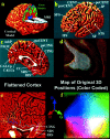
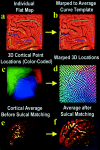
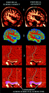
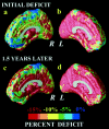
We detected and mapped a dynamically spreading wave of gray matter loss in the brains of patients with Alzheimer's disease (AD). The loss pattern was visualized in four dimensions as it spread over time from temporal and limbic cortices into frontal and occipital brain regions, sparing sensorimotor cortices. The shifting deficits were asymmetric (left hemisphere > right hemisphere) and correlated with progressively declining cognitive status (p < 0.0006). Novel brain mapping methods allowed us to visualize dynamic patterns of atrophy in 52 high-resolution magnetic resonance image scans of 12 patients with AD (age 68.4 +/- 1.9 years) and 14 elderly matched controls (age 71.4 +/- 0.9 years) scanned longitudinally (two scans; interscan interval 2.1 +/- 0.4 years). A cortical pattern matching technique encoded changes in brain shape and tissue distribution across subjects and time. Cortical atrophy occurred in a well defined sequence as the disease progressed, mirroring the sequence of neurofibrillary tangle accumulation observed in cross sections at autopsy. Advancing deficits were visualized as dynamic maps that change over time. Frontal regions, spared early in the disease, showed pervasive deficits later (>15% loss). The maps distinguished different phases of AD and differentiated AD from normal aging. Local gray matter loss rates (5.3 +/- 2.3% per year in AD v 0.9 +/- 0.9% per year in controls) were faster in the left hemisphere (p < 0.029) than the right. Transient barriers to disease progression appeared at limbic/frontal boundaries. This degenerative sequence, observed in vivo as it developed, provides the first quantitative, dynamic visualization of cortical atrophic rates in normal elderly populations and in those with dementia.
Figures


References
-
- American Psychiatric Association. Diagnostic and statistical manual of mental disorders, Ed 4 (DSM-IV). American Psychiatric Association; Washington, DC: 2000.
-
- Arnold SE, Hyman BT, Flory J, Damasio AR, Van Hoesen GW. The topographical and neuroanatomical distribution of neurofibrillary tangles and neuritic plaques in the cerebral cortex of patients with Alzheimer's disease. Cereb Cortex. 1991;1:103–116. - PubMed
-
- Arriagada P, Growdon JH, Hedley-Whyte ET, Hyman BT. Neurofibrillary tangles, but not plaques parallel duration and severity of Alzheimer's disease. Neurology. 1992;42:631–639. - PubMed
-
- Ashburner J, Friston KJ. Voxel-based morphometry—the methods. NeuroImage. 2000;11:805–821. - PubMed
-
- Ashburner J, Friston KJ. Why voxel-based morphometry should be used. NeuroImage. 2001;14:1238–1243. - PubMed
Publication types
MeSH terms
Grants and funding
LinkOut - more resources
Full Text Sources
Other Literature Sources
Medical
