. 2003 Feb 18;100(4):1552-7.
doi: 10.1073/pnas.0437829100. Epub 2003 Feb 6.

The N-terminal domain of mammalian soluble epoxide hydrolase is a phosphatase

Affiliations

The N-terminal domain of mammalian soluble epoxide hydrolase is a phosphatase

Annette Cronin et al. Proc Natl Acad Sci U S A. .

Abstract

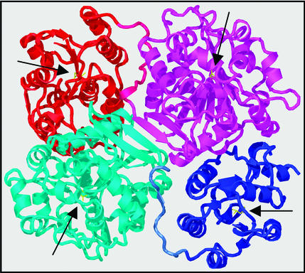
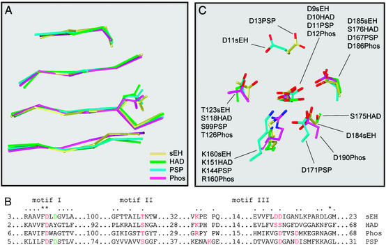
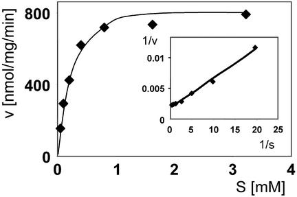
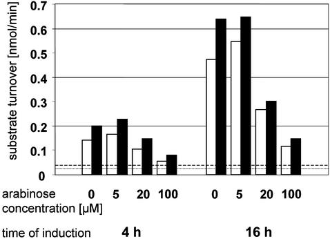
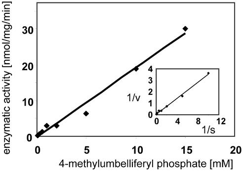
The mammalian soluble epoxide hydrolase (sEH) is an enzyme with multiple functions, being implicated in detoxification of xenobiotic epoxides as well as in regulation of physiological processes such as blood pressure. The enzyme is a homodimer, in which each subunit is composed of two domains. The 35-kDa C-terminal domain has an alpha/beta hydrolase fold and harbors the catalytic center for the EH activity. The 25-kDa N-terminal domain has a different alpha/beta fold and belongs to the haloacid dehalogenase superfamily of enzymes. The catalytic properties of the enzyme reported so far can all be explained by the action of the C-terminal domain alone. The function of the N-terminal domain, other than in structural stabilization of the dimer, has therefore remained unclear. By structural comparison of this domain to other haloacid dehalogenase family members, we identified a putative active site containing all necessary components for phosphatase activity. Subsequently, we found rat sEH hydrolyzed 4-nitrophenyl phosphate with a rate constant of 0.8 s(-1) and a K(m) of 0.24 mM. Recombinant human sEH lacking the C-terminal domain also displayed phosphatase activity. Presence of a phosphatase substrate did not affect epoxide turnover nor did epoxides affect dephosphorylation by the intact enzyme, indicating both catalytic sites act independently. The enzyme was unable to hydrolyze 4-nitrophenyl sulfate, suggesting its role in xenobiotic metabolism does not extend beyond phosphates. Thus, we propose this domain participates instead in the regulation of the physiological functions associated with sEH.


Figures

Figure 1
Figure 1
3D structure of mammalian sEH. The sEH structure is displayed as a cartoon prepared by using the program O (21). Coordinates were taken from the Protein Data Bank (ID code 1cqz) (17). The N- and C-terminal domains of one subunit are colored in blue and cyan, respectively, whereas they are red and magenta in the other subunit. The side chains of the catalytic nucleophiles of all four active sites (see text) are shown in yellow. Their respective positions are indicated by black arrows.
Figure 2
Figure 2
Structural/sequence alignment of mammalian sEH with haloacid dehalogenase, phosphonoacetaldehyde hydrolase, and phosphoserine phosphatase. Based on the DALI results (see text), four structures were superimposed. (A) The alignment of the four central strands of the β sheet, in the order β2, -1, -4, and -5 from top to bottom [strand designation according to Morais et al. (28)], including the catalytic nucleophile sidechain. (B) A structure-based sequence alignment in the vicinity of (potential) catalytic residues, which are marked in red. Designation of motifs is adopted from Wang et al. (30), with the numbers of residues in the linking segments given for each sequence. (C) The spatial arrangement of these residues. Phos, phosphonoacetaldehyde hydrolase; PSP, phosphoserine phosphatase. Protein Data Bank entries are 1cqz (sEH), 1jud (HAD), and 1fez (Phos). 1f5s (PSP). The spatial arrangement observed for PSP has also been seen more recently in the structure from another phosphatase from the HAD family (38).
Figure 3
Figure 3
4-Nitrophenyl phosphate hydrolysis by native rat sEH. Concentration-dependent 4-nitrophenol dephosphorylation is shown in the Michaelis–Menten representation. (Inset) The corresponding Lineweaver–Burk plot.
Figure 4
Figure 4
Recombinant expression of the full-length rat sEH in E. coli BL21-AI. The bar graph shows the relationship between the concentration of the inducer arabinose and the yield of enzymatically active sEH. The open bars represent the hydrolysis of trans-stilbene oxide obtained with 60 μg of protein from the bacterial lysate supernatant, whereas the filled bars show the 4-nitrophenyl phosphate hydrolysis with 40 μg of the same material. Constitutive phosphatase activity levels and spontaneous transstilbene oxide hydrolysis levels in sEH-free E. coli lysates are indicated by broken and dotted lines, respectively. Notably, the EH activity is perfectly paralleled by the phosphatase activity. The production of active enzyme is maximal at an arabinose concentration of 5 μM. Beyond that, enhanced inclusion body formation leads to a disproportionate increase in the amount of improperly folded recombinant sEH, and the yield in active sEH drops substantially.
Figure 5
Figure 5
Enzyme kinetics of 4-methylumbelliferyl phosphate hydrolysis by rat sEH. Displayed is the dependence of the turnover rate on the substrate concentration. Note the lack of any indication of saturation. Therefore, the rate constant under saturating conditions cannot be determined. However, kcat/Km can be obtained by mathematical transformation of the slope of the Lineweaver–Burk plot (Inset), which itself represents Km/Vmax.

References

    1. Arand M, Oesch F. In: Enzyme Systems That Metabolise Drugs and Other Xenobiotics. Ioannides C, editor. New York: Wiley; 2002. pp. 459–483.
    1. Oesch F. Xenobiotica. 1973;3:305–340. - PubMed
    1. Moghaddam M F, Grant D F, Cheek J M, Greene J F, Williamson K C, Hammock B D. Nat Med. 1997;3:562–566. - PMC - PubMed
    1. Yu Z, Xu F, Huse L M, Morisseau C, Draper A J, Newman J W, Parker C, Graham L, Engler M M, Hammock B D, et al. Circ Res. 2000;87:992–998. - PubMed
    1. Sinal C J, Miyata M, Tohkin M, Nagata K, Bend J R, Gonzalez F J. J Biol Chem. 2000;275:40504–40510. - PubMed

Publication types

MeSH terms