. 2003 Feb 18;100(4):1996-2001.
doi: 10.1073/pnas.0337370100. Epub 2003 Feb 6.

Genome-wide protective response used by group A Streptococcus to evade destruction by human polymorphonuclear leukocytes

Affiliations

Genome-wide protective response used by group A Streptococcus to evade destruction by human polymorphonuclear leukocytes

Jovanka M Voyich et al. Proc Natl Acad Sci U S A. .

Abstract

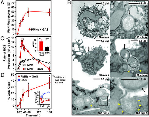
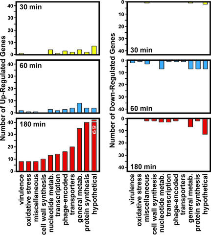
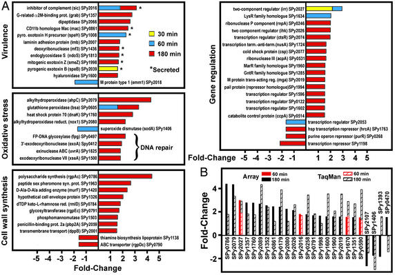
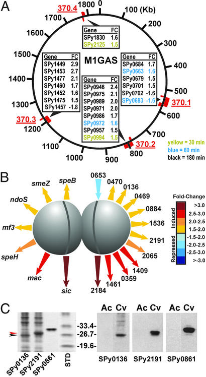
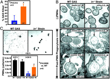
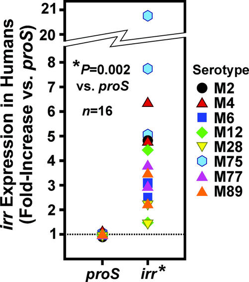
Group A Streptococcus (GAS) evades polymorphonuclear leukocyte (PMN) phagocytosis and killing to cause human disease, including pharyngitis and necrotizing fasciitis (flesh-eating syndrome). We show that GAS genes differentially regulated during phagocytic interaction with human PMNs comprise a global pathogen-protective response to innate immunity. GAS prophage genes and genes involved in virulence, oxidative stress, cell wall biosynthesis, and gene regulation were up-regulated during PMN phagocytosis. Genes encoding novel secreted proteins were up-regulated, and the proteins were produced during human GAS infections. We discovered an essential role for the Ihk-Irr two-component regulatory system in evading PMN-mediated killing and promoting host-cell lysis, processes that would facilitate GAS pathogenesis. Importantly, the irr gene was highly expressed during human GAS pharyngitis. We conclude that a complex pathogen genetic program circumvents human innate immunity to promote disease. The gene regulatory program revealed by our studies identifies previously undescribed potential vaccine antigens and targets for therapeutic interventions designed to control GAS infections.


Figures

Figure 1
Figure 1
GAS–PMN interaction. (A) Phagocytosis of GAS. Phagocytosis (%) is the percent of PMNs that contain ingested GAS at each time. Results are the mean ± SD of three experiments. (B) Ultrastructural analyses of PMNs containing ingested GAS. Black arrows indicate ingested GAS. (Right) Higher magnification. Yellow arrowheads indicate fusion of PMN granules with GAS phagosomes. (C) ROS production during phagocytosis of GAS. Data shown are from a representative experiment performed four times. Inset illustrates the rate of ROS production at Vmax (†) (mean ± SD of four experiments). *, P = 0.006 vs. unstimulated PMNs. (D) Killing of GAS by PMNs. At each time, PMNs were lysed and GAS were plated on Todd–Hewitt agar containing yeast extract. Colonies were enumerated the following day, and percent GAS killed was calculated by using the equation (1 − (CFUPMN+/CFUPMN−)) × 100 as described in Supporting Methods. Results are the mean ± SD of three experiments. *, P < 0.03 vs. PMNs + GAS at 0 min, one-way ANOVA with Tukey posttest. Inset illustrates representative growth curves of GAS in the presence (red) and absence (blue) of PMNs.
Figure 2
Figure 2
Differential GAS gene expression during PMN phagocytosis. PMNs were incubated with GAS for the indicated times, and gene expression was measured with a DNA microarray containing 1,705 (of 1,752) serotype M1 GAS ORFs, 97.3% of the genome. Data were analyzed with genespring software, version 4.2 (Silicon Genetics).
Figure 3
Figure 3
GAS genome-wide protective response to PMN phagocytosis. (A) GAS genes differentially expressed during PMN phagocytosis. Results are presented as the mean fold-induction or repression of genes from three experiments (three blood donors with phagocytosis assays done on separate days) and three accompanying microarray experiments (done on separate days). (B) TaqMan confirmation of microarray results. Genes (n = 26) identified as differentially transcribed by GAS microarrays were analyzed by TaqMan real-time PCR. There was a strong positive correlation (r = 0.76) between TaqMan and microarray results, consistent with previous comparisons (15, 25).
Figure 4
Figure 4
Genes encoding GAS prophage and secreted proteins. (A) Prophage-encoded genes differentially regulated during phagocytosis. Genes were assigned to prophage (red) based on homology with serotype M1 GAS SF370 (11). (B) Differential expression of genes encoding secreted proteins of known (left) and unknown (right) function. Length of arrows is relative for gene expression. (C) Production of novel secreted proteins in humans with GAS disease. SPy0136 and SPy2191 were overexpressed in E. coli and separated by SDS-PAGE (Gel-Code protein stain, Left). Comparison of acute (Ac) and convalescent (Cv) paired sera from an individual with GAS invasive disease (Right). Data are representative of paired human sera from three individuals with GAS invasive disease.
Figure 5
Figure 5
Ihk-Irr two-component gene regulatory system is essential for GAS survival and host-cell lysis. (A) Killing of mutant (irr) strain by PMNs at 180 min. Results are the mean ± SD of three experiments. (B) Ultrastructural analyses of PMNs containing ingested wild-type (WT) or mutant (irr) strains at 180 min. Black arrows indicate ingested GAS. (C) Isogenic GAS mutant (irr) strain has reduced ability to cause PMN lysis. Results are the mean ± SD of three to five experiments.
Figure 6
Figure 6
The irr gene is highly expressed in vivo in human infections. Transcript levels for irr were determined by TaqMan real-time PCR in 16 patients with GAS pharyngitis. Results are expressed as fold-increase compared with the GAS proS gene.

References

    1. Jones S L, Lindberg F P, Brown E J. In: Fundamental Immunology. Paul W E, editor. New York: Lippincott-Raven; 1999. pp. 997–1021.
    1. Nauseef W M, Clark R A. In: Basic Principles in the Diagnosis and Management of Infectious Diseases. Mandell G L, Bennett J E, Dolin R, editors. New York: Churchill Livingstone; 2000. pp. 89–112.
    1. Cunningham M W. Clin Microbiol Rev. 2000;13:470–511. - PMC - PubMed
    1. Frank M M. Nat Med. 2001;7:1285–1286. - PubMed
    1. Lei B, DeLeo F R, Hoe N P, Graham M R, Mackie S M, Cole R L, Liu M, Hill H R, Low D E, Federle M J, et al. Nat Med. 2001;7:1298–1305. - PubMed