. 2003 Mar 4;100(5):2231-6.
doi: 10.1073/pnas.0336132100. Epub 2003 Feb 6.

Cloning, expression, and characterization of a membrane progestin receptor and evidence it is an intermediary in meiotic maturation of fish oocytes

Affiliations

Cloning, expression, and characterization of a membrane progestin receptor and evidence it is an intermediary in meiotic maturation of fish oocytes

Yong Zhu et al. Proc Natl Acad Sci U S A. .

Abstract

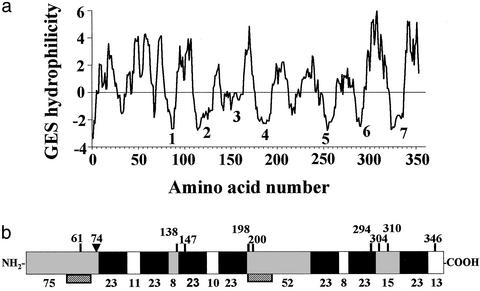
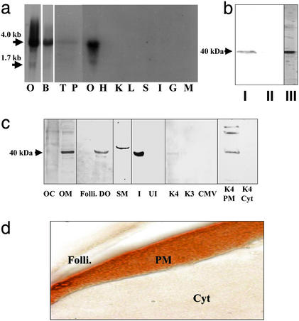
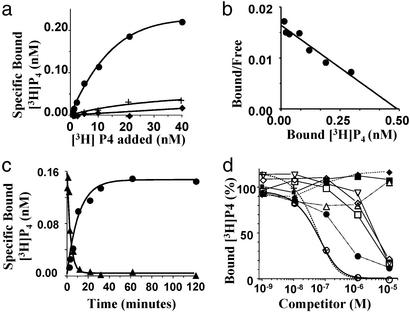
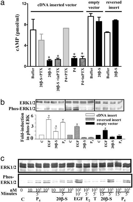
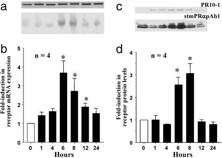
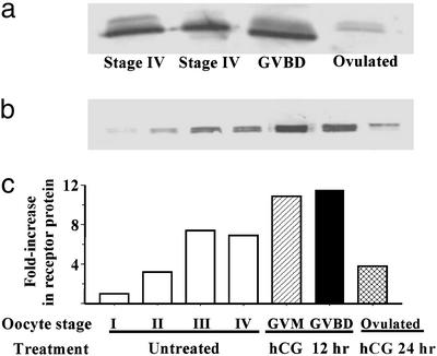
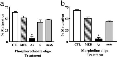
The structures of membrane receptors mediating rapid, nongenomic actions of steroids have not been identified. We describe the cloning of a cDNA from spotted seatrout ovaries encoding a protein that satisfies the following seven criteria for its designation as a steroid membrane receptor: plausible structure, tissue specificity, cellular distribution, steroid binding, signal transduction, hormonal regulation, and biological relevance. For plausible structure, computer modeling predicts that the protein has seven transmembrane domains, typical of G protein-coupled receptors. The mRNA (4.0 kb) is only detected in the brain and reproductive tissues on Northern blots. Antisera only detect the protein (40 kDa) in plasma membranes of reproductive tissues. The recombinant protein produced in an Escherichia coli expression system has a high affinity (K(d) = 30 nM), saturable, displaceable, single binding site specific for progestins. Progestins alter signal transduction pathways, activating mitogen-activated protein kinase and inhibiting adenylyl cyclase, in a transfected mammalian cell line. Inhibition of adenylyl cyclase is pertussis toxin sensitive, suggesting the receptor may be coupled to an inhibitory G protein. Progestins and gonadotropin up-regulate both mRNA and protein levels in seatrout ovaries. Changes in receptor abundance in response to hormones and at various stages of oocyte development, its probable coupling to an inhibitory G protein and inhibition of progestin induction of oocyte maturation upon microinjection of antisense oligonucleotides are consistent with the identity of the receptor as an intermediary in oocyte maturation. These characteristics suggest the fish protein is a membrane progestin receptor mediating a "nonclassical" action of progestins to induce oocyte maturation in fish.


Figures

Figure 1
Figure 1
Structural analysis of deduced amino acid sequence of putative mPR gene. (a) Hydrophilicity profile according to GES method (Goldman/Engelman/Steitz, 1986). (b) Schematic diagram of encoded protein showing extracellular (gray), seven-transmembrane (solid black), and cytoplasmic (clear) domains predicted by several programs. Vertical lines indicate potential phosphorylation sites. ▾, potential N-linked glycosylation site. Numbers above: amino acids from N-terminal end; numbers below: number of amino acids in each domain. Hatched boxes: peptide sequences used to generate polyclonal antibodies.
Figure 2
Figure 2
Tissue distribution and cellular localization of putative mPR. (a) Northern blot analysis showing mRNA expression in ovarian (O, gel loading: 1 μg) and other tissues (gel loading: 5 μg); B, brain; T, testis; P, pituitary; H, heart; K, kidney; L, liver; S, stomach; I, intestine; G, gill; M, muscle. (b) Western blot analysis of solubilized ovarian membrane proteins using monoclonal antibody PR10-1. I, plasma membrane; II, cytosol; III, partially purified membrane fraction used to generate monoclonal antibodies. (c) Cellular localization of receptor by Western blot analysis using stmPRαpAb1 antibody (gel loading: 10 μg). OC, oocyte cytosol; OM, oocyte membrane; Folli., follicle cell membrane; DO, denuded oocyte plasma membrane; SM, sperm membrane; I, recombinant protein induced by IPTG in E. coli; UI, noninduced E. coli protein; K4, membrane proteins from mPR-transfected MDA-MB-231 cells; K3, control cells transfected with vector containing reverse insert; CMV, control cells transfected with empty carrier vector. The following lanes were probed with PR10-1 antibody: K4 PM, plasma membrane from mPR transfected MDA-MB-231 cells; K4 Cyt, cytosol from mPR transfected cells. (d) Immunocytochemical localization of mPR receptor protein in a mature seatrout follicle containing a stage IV oocyte using stmPRαpAb1 antibody. Folli., follicle cells; PM, oocyte plasma membrane; Cyt, oocyte cytoplasm. (Magnification: 1 cm ≈ 20 μm.)
Figure 3
Figure 3
Steroid binding characteristics of recombinant mPR protein produced in E. coli. (a) Representative plots of specific [3H]progesterone binding to protein produced with a cDNA-inserted vector with (●) or without (♦) IPTG induction, or an empty carrier vector (+, controls). (b) Scatchard analysis of specific [3H]progesterone binding. (c) Time course of association and dissociation of specific [3H]progesterone binding. (d) Competition curves of steroid binding expressed as a percentage of maximum specific [3H]progesterone binding [concentration: [3H]progesterone, 10 nM, specific activity; 65 Ci/mmol (1 Ci = 37 GBq); Amersham Pharmacia]; ○, progesterone; +, 17-hydroxyprogesterone; ●, 20β-hydroxyprogesterone; ▵, 20β-S; □, testosterone; ⋄, 17,20β-dihydroxy-4-pregnen-3-one; ▿, 11-deoxycortisol; ♦, estradiol-17β; ■, cortisol.
Figure 4
Figure 4
Inhibition of cAMP production (a) and activation of MAP kinase signaling pathway (b and c) in response to progestin hormones in MDA-MB-231 cells stably transfected with putative mPR cDNA (means ± SEM, n = 4; *, P < 0.05). (a) Cells were preexposed to activated pertussis toxin (PTX) or inactive pertussis toxin (inPTX) for 30 min at 37°C before incubation with 20β-S or progesterone for 5 min. (b) MAP kinase activity (shown as increase in phospho-Erk1/2) in cells transfected with vector containing mPR insert (white bar), controls containing reversed insert (shaded bar) or vector alone (black bar) after a 5-min stimulation with 1 μM 20β-S or progesterone (P4). C, untreated control; EGF, human epidermal growth factor (1 μM, positive control). (c) Time- and dose-dependent activation of MAP kinase (gel loading: 20 μg per lane) in transfected cells treated with 20β-S, P4, estradiol-17β (E2), or testosterone (T).
Figure 5
Figure 5
Time course of changes in putative mPR mRNA (a and b) and protein levels (c and d) in ovarian fragments after in vitro treatment with 300 nM 20β-S, relative to control (0 h) values (mean ± SEM, n = 4; *, P < 0.05). (a) Representative Northern blot (gel loading: 1 μg). (Lower) Putative mPR mRNA. (Upper) Corresponding β-actin mRNA. (b) Relative levels of mPR mRNA. (c) Representative Western blot analyses using PR10-1 and stmPRαAb1 antibodies (gel loading: 10 μg). (d) Relative levels of mPR protein.
Figure 6
Figure 6
Changes in putative mPR protein levels in oocytes at various stages of development and final maturation detected by Western blot analysis with stmPRαAb1 antibody (gel loading: membrane proteins from five oocytes per lane). (a) In wild fish captured on their spawning grounds. (b) After incubation in vitro in the presence or absence of hCG. I, vitellogenic oocyte, 200–300 μm (diameter); II, 300–375 μm; III, 375–440 μm; IV, full-grown oocyte, 430–500 μm; GVM, germinal vesicle migration stage of final maturation; GVBD, germinal vesicle breakdown stage of final maturation. (c) mPR protein levels expressed relative to stage I oocyte values.
Figure 7
Figure 7
Effects of microinjection of zebrafish oocytes with zebrafish antisense oligonucleotides on their subsequent maturation in response to 17,20β-P (mean ± SEM, n = 4; *, P < 0.05). (a) Phosphorothioate antisense experiment. CTL, control uninjected oocytes; MED, injected with media alone; As, zebrafish mPR α-antisense; S, mPR α-sense; mAS, mPR α-mis-antisense. (b) Morpholino antisense experiment.

Comment in

References

    1. Yang J, Serres C, Philibert D, Robel P, Baulieu E-E, Jouannet P. Proc Natl Acad Sci USA. 1994;91:529–533. - PMC - PubMed
    1. Lieberherr M, Grosse B. J Biol Chem. 1994;269:7217–7223. - PubMed
    1. Gu Q, Korach K S, Moss R L. Endocrinology. 1999;140:660–666. - PubMed
    1. Pietras R, Szego C. Nature. 1975;253:357–359. - PubMed
    1. Qui J, Lou G, Huang Y, Lou J, Chen Z. Endocrinology. 1998;139:5103–5108. - PubMed

Publication types

MeSH terms