. 2003 Mar 1;547(Pt 2):333-48.
doi: 10.1113/jphysiol.2002.034140. Epub 2003 Feb 7.

Store-operated Ca2+ entry: dynamic interplay between endoplasmic reticulum, mitochondria and plasma membrane

Affiliations

Store-operated Ca2+ entry: dynamic interplay between endoplasmic reticulum, mitochondria and plasma membrane

Anant B Parekh. J Physiol. .

Abstract

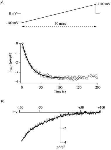
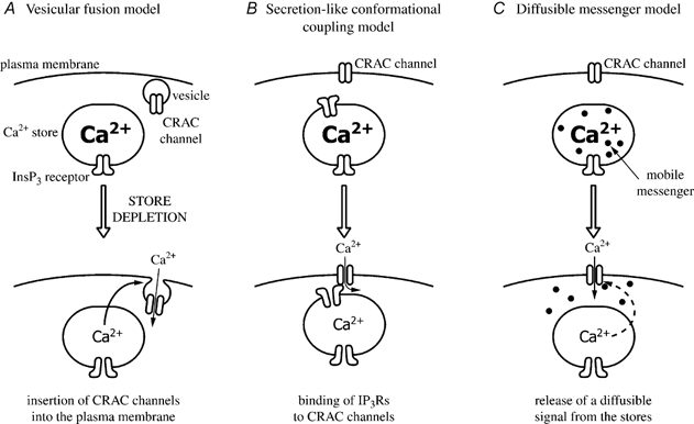
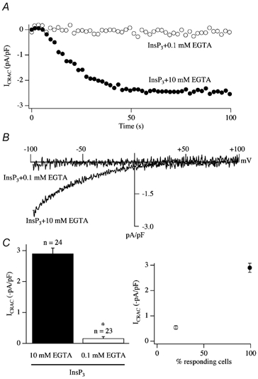
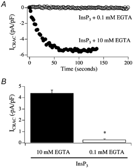
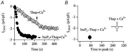
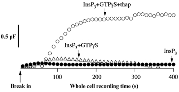
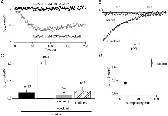
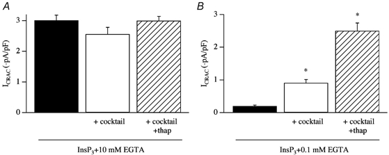
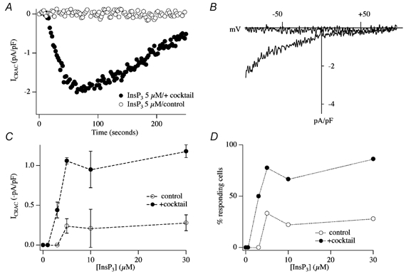
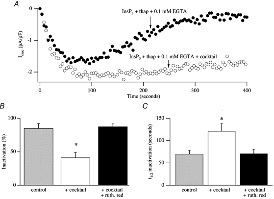
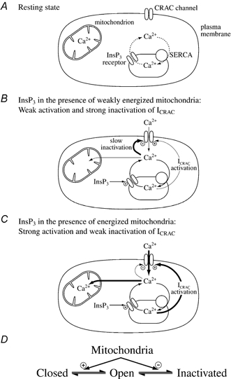
In eukaryotic cells, hormones and neurotransmitters that engage the phosphoinositide pathway evoke a biphasic increase in intracellular free Ca2+ concentration: an initial transient release of Ca2+ from intracellular stores is followed by a sustained phase of Ca2+ influx. This influx is generally store-dependent and is required for controlling a host of Ca2+-dependent processes ranging from exocytosis to cell growth and proliferation. In many cell types, store-operated Ca2+ entry is manifest as a non-voltage-gated Ca2+ current called ICRAC (Ca2+ release-activated Ca2+ current). Just how store emptying activates CRAC channels remains unclear, and some of our recent experiments that address this issue will be described. No less important from a physiological perspective is the weak Ca2+ buffer paradox: whereas macroscopic (whole cell) ICRAC can be measured routinely in the presence of strong intracellular Ca2+ buffer, the current is generally not detectable under physiological conditions of weak buffering following store emptying with the second messenger InsP3. In this review, I describe some of our experiments aimed at understanding just why InsP3 is ineffective under these conditions and which lead us to conclude that respiring mitochondria are essential for the activation of ICRAC in weak intracellular Ca2+ buffer. Mitochondrial Ca2+ uptake also increases the dynamic range over which InsP3 functions as the second messenger that controls Ca2+ influx. Finally, we find that Ca2+-dependent slow inactivation of Ca2+ influx, a widespread but poorly understood phenomenon that helps shape the profile of an intracellular Ca2+ signal, is regulated by mitochondrial Ca2+ buffering. Thus, by enabling macroscopic store-operated Ca2+ current to activate and then by controlling its extent and duration, mitochondria play a crucial role in all stages of store-operated Ca2+ influx. Store-operated Ca2+ entry reflects therefore a dynamic interplay between endoplasmic reticulum, mitochondria and plasma membrane.


Figures

Figure 1
Figure 1. Development of ICRAC following store depletion
A, the upper panel shows the typical voltage ramp protocol used to monitor ICRAC in mast cells, the related rat basophilic leukaemia (RBL-1) cell line and jurkat T-lymphocytes. The ramp spans −100 to +100 mV in 50 ms, and is applied from a holding potential of 0 mV once every 2 s. The lower panel depicts the time course of activation of ICRAC. The current amplitude, measured at −80 mV from each ramp, has been normalised for cell size by dividing the current by cell capacitance. The thick line shows a mono-exponential fit to the activation time course. B shows the current-voltage relationship, once the current had reached steady state. Note the inward rectification and very positive reversal potential, characteristic of ICRAC. The pipette solution is caesium glutamate based, supplemented with InsP3 and 10 mm EGTA.
Figure 2
Figure 2. The three contemporary models proposed to account for the activation mechanism of ICRAC
Figure 3
Figure 3. The weak buffer paradox
A, InsP3 generally fails to activate ICRAC in weak intracellular Ca2+ buffer (0.1 mm EGTA) whereas it is effective in strong buffer (10 mm EGTA). B, current-voltage relationships for the two recordings from A, taken at steady state. C, the mean amplitude of ICRAC (left-hand panel) and the fraction of cells responding (right-hand panel) for each condition is shown. In this and all subsequent figures, data from RBL-1 cells are shown. *P < 0.01.
Figure 4
Figure 4. InsP3 still fails to activate ICRAC in weak Ca2+ buffer at physiological temperature
Experiments were carried out at 36 °C. A, whereas InsP3 evoked a large ICRAC at 36 °C in 10 mm EGTA, no current was detectable in 0.1 mm EGTA. B, aggregate data are summarised. *P < 0.01 (Student's t test).
Figure 5
Figure 5. Passive activation of ICRAC
A, comparison of the time course of development of ICRAC following dialysis with either 10 mm EGTA alone or 30 μm InsP3+ EGTA. Note the initial slow development of ICRAC followed by a second, faster phase in response to EGTA alone. B, development of ICRAC following dialysis with 10 mm BAPTA or dimethyl BAPTA (panel C).
Figure 6
Figure 6. SERCA pumps shape the pattern of passive activation of ICRAC
A, inclusion of cyclopiazonic acid (CPA) eliminates the initial slow phase of current development, leaving the second component intact. B, following dialysis with 2.5 mm EGTA, ICRAC activated partially in this cell. Subsequent application of thapsigargin increased the extent of the current, as well as its rate of development.
Figure 7
Figure 7. SERCA pumps function in the presence of InsP3
A, the relationship between intrapipette EGTA concentration and amplitude of ICRAC (evoked by dialysis with 30 μm InsP3) is plotted. Note that ICRAC was clearly submaximal in the presence of 0.6 mm EGTA. Each point is the mean ±s.e.m. of at least 5 cells. B, recordings are shown for a cell dialysed with 30 μm InsP3 and 0.6 mm EGTA (•) and for one dialysed with this solution but supplemented with 2 μm thapsigargin, a SERCA blocker (○). Note the increase in the size of ICRAC following inhibition of the pumps. C, the amplitudes, delays and times to peak are summarised for experiments as in B. Filled circles denote InsP3+ 0.6 mm EGTA whereas open cirlces represent InsP3+ 0.6 mm EGTA + thapsigargin.
Figure 8
Figure 8. InsP3 activates ICRAC in weak buffer provided SERCA pump activity is compromised
A, InsP3 together with thapsigargin activates ICRAC in weak buffer whereas InsP3 alone is ineffective. B, InsP3 with cyclopiazonic acid, a structurally distinct SERCA pump blocker, is also effective. SERCA pump block alone (2 μm Thap + 0.1 mm EGTA) can also activate ICRAC, albeit at a much slower rate.
Figure 9
Figure 9. SERCA pump block is sufficient to activate ICRAC even in high intracellular Ca2+
A, InsP3 and thapsigargin can activate ICRAC in weak buffer even in the presence of high intracellular Ca2+ (100 μm total CaCl2 in the pipette solution, free Ca2+ concentration estimated to be in the micromolar range). Thapsigargin alone was also able to evoke ICRAC under these conditions, but after a delay and at a slower rate. B, aggregate data are summarised.
Figure 10
Figure 10. The combination of InsP3 and thapsigargin evokes a robust secretory response in weak buffer, whereas InsP3 alone is largely ineffective
InsP3 alone (•) or in combination with GTPγS (▵) did not trigger a detectable increase in membrane capacitance, whereas InsP3 together with thapsigargin and GTPγS produced a prominent secretory response (○). In these experiments, cells were clamped at −60 mV.
Figure 11
Figure 11. InsP3 activates ICRAC in weak buffer provided mitochondria are energised
A, time course of activation of ICRAC in the presence of the mitochondrial cocktail (○). ICRAC does not activate in weak Ca2+ buffer in response to InsP3 alone (•). B, steady-state I-V relationships are shown for the cells in A. C, the effects of cocktail are suppressed by preventing mitochondrial Ca2+ uptake by either depolarising mitochondria (pre-treatment with antimycin A (5 μg ml−1) and oligomycin (0.5 μg ml−1)) or inhibiting the uniporter with ruthenium red. D, the presence of cocktail dramatically increases the fraction of cells that respond to InsP3 in weak Ca2+ buffer. *P < 0.01.
Figure 12
Figure 12. Physiological antagonism between SERCA pumps and mitochondria
A, the potentiation of ICRAC by cocktail is suppressed by strong intracellular Ca2+ buffer (10 mm EGTA), indicating that the effect is Ca2+-dependent. B, the extent of ICRAC to InsP3 in weak buffer alone, in mitochondrial cocktail and cocktail plus thapsigargin is compared. *P < 0.01.
Figure 13
Figure 13. Energised mitochondria increase the ability of InsP3 to activate ICRAC in weak Ca2+ buffer
A, whereas 5 μm InsP3 is ineffective in weak buffer (○), it evokes a prominent ICRAC in the presence of energised mitochondria (•). B, I-V relationships for the cells shown in A, taken at steady state. C, concentration-response curve to InsP3 in the absence (○) and presence (•) of cocktail. D, the fraction of cells responding versus concentration of InsP3 is plotted in the absence (○) and presence (•) of energised mitochondria.
Figure 14
Figure 14. Mitochondrial Ca2+ uptake reduces the rate and extent of Ca2+-dependent slow inactivation
A, dialysis with InsP3 and thapsigargin in weak buffer (control) activates ICRAC but then the current inactivates slowly (•). The extent of inactivation is reduced if mitochondria are energised (○). B and C summarise aggregate data comparing the extent (B) and rate (C) of inactivation of ICRAC in control cells, in those in which cocktail was added to the solution and in cells exposed to cocktail and ruthenium red, to inhibit mitochondrial Ca2+ uptake. *P < 0.01. t1/2, time at which ICRAC had inactivated by 50 %.
Figure 15
Figure 15. A diagrammatic scheme for the role of mitochondria in controlling ICRAC under physiological conditions of weak intracellular Ca2+ buffering
A, the resting state, where ICRAC is not functioning, is shown. Stores are largely full and any Ca2+ that leaks from the stores is taken back up by the SERCA pumps. B, increasing InsP3 levels in the absence of active mitochondrial Ca2+ uptake, releases Ca2+ from the stores. However, the SERCA pumps are able to resequester sufficient Ca2+ so only a very small fraction of CRAC channels are activated (undetectable in whole cell mode). Furthermore, the rise in cytosolic Ca2+ results in Ca2+-dependent slow inactivation of CRAC channels, and possibly InsP3 receptors as well. C, in the presence of respiring mitochondria, InsP3 activates macroscopic ICRAC. Ca2+ released from the stores by InsP3 is taken up by mitochondria. This reduces the amount of Ca2+ available to the SERCA pumps and in the vicinity of open InsP3 receptors such that the stores are depleted sufficiently for macroscopic ICRAC to activate (less refilling by SERCA pumps and less inactivation of InsP3 receptors). Some refilling does occur because inclusion of thapsigargin enhances the size of the current. Furthermore, mitochondrial Ca2+ buffering reduces the rate and extent of Ca2+-dependent slow inactivation, thereby increasing the size and duration of the current. D, a simplified gating scheme for CRAC channels summarising the role of mitochondrial Ca2+ buffering. Mitochondria facilitate opening (Closed to Open transition) whilst, simultaneously, reducing inactivation (Open to Inactivated transition). In this way, mitochondria have a much larger impact on ICRAC than through either transition alone.

References

    1. Alderton JM, Ahmed SA, Smith LA, Steinhardt RA. Evidence for a vesicle-mediated maintenance of store-operated calcium channels in a human embryonic kidney cell line. Cell Calcium. 2000;28:161–169. - PubMed
    1. Arnaudeau S, Kelley WL, Walsh JV, Demaurex N. Mitochondria recycle Ca2+ to the endoplasmic reticulum and prevent the depletion of neighbouring endoplasmic reticulum regions. J Biol Chem. 2001;276:29430–29439. - PubMed
    1. Artalejo AR, Ellory JC, Parekh AB. Ca2+ -dependent capacitance increases in rat basophilic leukemia cells following activation of store-operated Ca2+ entry and dialysis with high-Ca2+-containing intracellular solution. Pflugers Arch. 1998;436:934–939. - PubMed
    1. Babcock DF, Herrington J, Goodwin PC, Park YB, Hille B. Mitochondrial participation in the intracellular Ca2+ network. J Cell Biol. 1997;136:833–844. - PMC - PubMed
    1. Bakowski D, Glitsch MD, Parekh AB. An examination of the secretion-like coupling model for the activation of the Ca2+ release-activated Ca2+ current ICRAC in RBl-1 cells. J Physiol. 2001;532:55–71. - PMC - PubMed

Publication types

LinkOut - more resources